 Back
BackCell Junctions: Structure, Function, and Genetic Relevance
Study Guide - Smart Notes
 Tailored notes based on your materials, expanded with key definitions, examples, and context.
Cell Junctions
Overview of Cell Junctions
Cell junctions are specialized structures that connect neighboring cells or cells to the extracellular matrix (ECM), enabling communication, adhesion, and coordination within tissues. These junctions are essential for maintaining tissue integrity and regulating cellular behavior.
Intercellular junctions: Connect cells to each other.
Cell-ECM junctions: Connect cells to the extracellular matrix.
Functions: Coordinate cell behavior, facilitate communication, and maintain tissue structure.
Types of Cell Junctions
Communicating junctions: Allow exchange of molecules between cells (e.g., gap junctions in animals, plasmodesmata in plants).
Occluding junctions: Prevent passage of molecules between cells (e.g., tight junctions).
Anchoring junctions: Provide mechanical stability by connecting cytoskeletal elements (e.g., desmosomes, adherens junctions, hemidesmosomes, focal adhesions).
Communicating Junctions
Gap Junctions (Animal Cells)
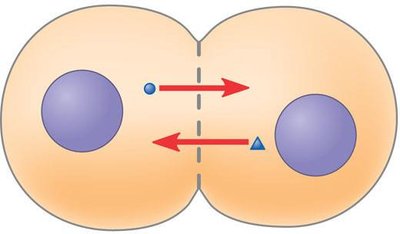
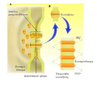
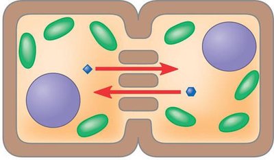
Gap junctions are specialized channels formed by connexin proteins that directly connect the cytoplasm of adjacent animal cells, allowing the passage of ions and small molecules.
Structure: Composed of connexin subunits forming a channel between cells.
Function: Enable cell-to-cell communication and synchronized activity (e.g., heart muscle contraction).
Examples: Transport of Ca2+ ions between smooth muscle cells for coordinated contraction.
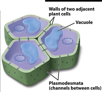
Plasmodesmata (Plant Cells)
Plasmodesmata are channels that traverse plant cell walls, connecting the cytoplasm of neighboring plant cells and facilitating the exchange of water, solutes, and macromolecules.
Structure: Channels through cell walls, lined by plasma membrane.
Function: Allow direct communication and transport between plant cells.
Examples: Movement of nutrients and signaling molecules in plant tissues.
Occluding Junctions
Tight Junctions
Tight junctions are occluding junctions found beneath the apical surface of epithelial cells. They create a barrier that prevents the passage of molecules between cells, maintaining distinct environments on either side of the epithelial layer.
Structure: Formed by claudin and occludin proteins, linked to actin microfilaments.
Function: Prevent leakage of extracellular fluid and inhibit intercellular communication.
Examples: Blood-brain barrier, skin, intestinal epithelium.
Anchoring Junctions
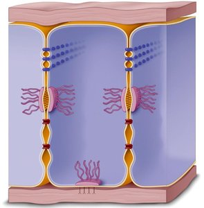
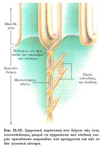
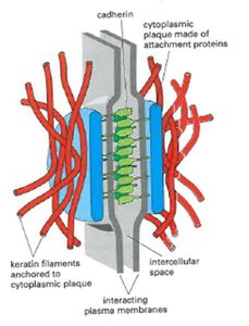
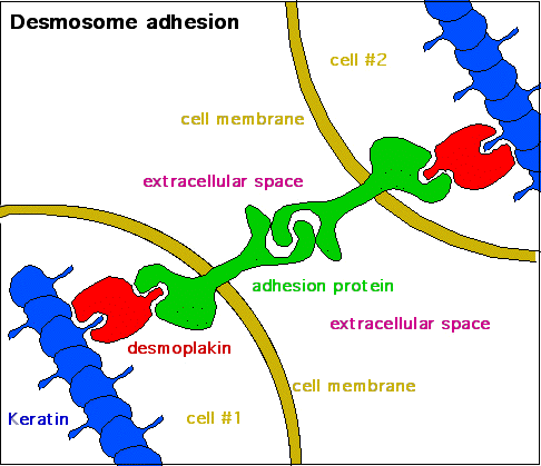
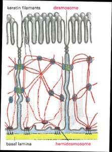
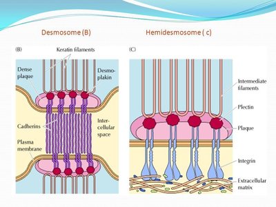
Desmosomes
Desmosomes are anchoring junctions that function like rivets, fastening cells together into strong sheets. They connect to intermediate filaments (e.g., keratin) via cadherin proteins, providing mechanical strength to tissues.
Structure: Cadherin proteins connect cells; attachment proteins (e.g., desmoplakin) link to intermediate filaments.
Function: Provide mechanical stability, especially in tissues subject to stress (e.g., skin, heart muscle).
Examples: Muscle tears can involve rupture of desmosomes.
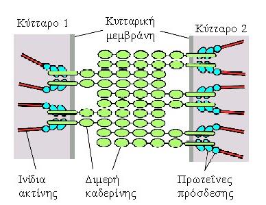
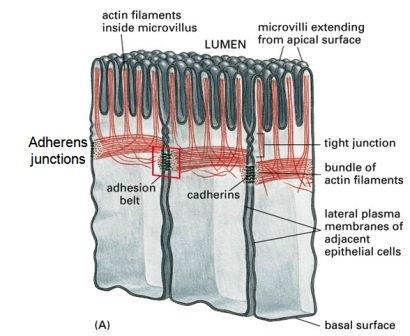
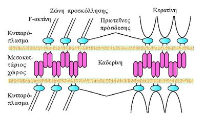
Adherens Junctions
Adherens junctions form adhesion belts beneath the apical surface of epithelial cells. They connect the plasma membranes of neighboring cells via cadherin proteins and link to actin microfilaments through intracellular attachment proteins.
Structure: Cadherin dimers connect cells; attachment proteins (catenins, vinculin, α-actinin) link to actin filaments.
Function: Maintain tissue integrity and facilitate cell shape changes.
Examples: Intestinal epithelial cells.
Comparison: Adherens Junctions vs Desmosomes
Both adherens junctions and desmosomes are anchoring junctions, but they differ in their cytoskeletal connections and structural proteins.
Adherens junctions: Connect to actin microfilaments via cadherins.
Desmosomes: Connect to intermediate filaments via cadherins and desmoplakin.
Cell-ECM Junctions: Focal Adhesions and Hemidesmosomes
Cell-ECM junctions anchor cells to the extracellular matrix, providing structural support and facilitating signal transduction.
Focal adhesions: Connect cells to ECM via integrins and actin microfilaments.
Hemidesmosomes: Anchor epithelial cells to the basement membrane via integrins and intermediate filaments (keratin).
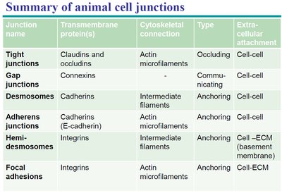
Summary Table: Animal Cell Junctions
The following table summarizes the main types of animal cell junctions, their structural proteins, cytoskeletal connections, and functions.
Junction name | Transmembrane protein(s) | Cytoskeletal connection | Type | Extracellular attachment |
|---|---|---|---|---|
Tight junctions | Claudins and occludins | Actin microfilaments | Occluding | Cell-cell |
Gap junctions | Connexins | - | Communicating | Cell-cell |
Desmosomes | Cadherins | Intermediate filaments | Anchoring | Cell-cell |
Adherens junctions | Cadherins (E-cadherin) | Actin microfilaments | Anchoring | Cell-cell |
Hemidesmosomes | Integrins | Intermediate filaments | Anchoring | Cell–ECM (basement membrane) |
Focal adhesions | Integrins | Actin microfilaments | Anchoring | Cell-ECM |
Clinical Correlations
Cell Junctions in Disease
Alterations in cell junctions are associated with various diseases:
Inflammation: Loss of tight junctions allows migration of immune cells.
Cancer: Changes in integrin expression facilitate metastasis.
Deafness: Mutations in connexin proteins can cause hearing loss.
Example Question
Which type of cell junction is only present in plant cells and not in animal cells?
A. Plasmodesmata
B. Tight junctions
C. Desmosomes
D. Gap junctions
E. Hemidesmosomes
Correct answer: A. Plasmodesmata
Additional info:
Cell junctions are critical for multicellular organization and genetic regulation of tissue development.
Mutations in junctional proteins can have profound effects on tissue function and organismal health.
