 Back
BackChapter 17: Recombinant DNA Technology – Study Notes
Study Guide - Smart Notes
 Tailored notes based on your materials, expanded with key definitions, examples, and context.
Ch 17 Recombinant DNA Technology
Introduction to Recombinant DNA Technology
Recombinant DNA technology, also known as gene splicing, involves the creation of DNA molecules by joining together DNA fragments from different sources. This technology allows scientists to isolate, study, and manipulate specific DNA sequences, enabling advances in genetics, biotechnology, and medicine. Clones are recovered copies of recombinant DNA molecules, used to study the structure and function of genes.
Tools of Recombinant DNA Technology
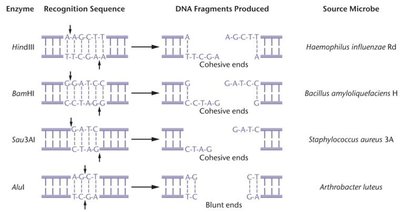
Restriction Enzymes
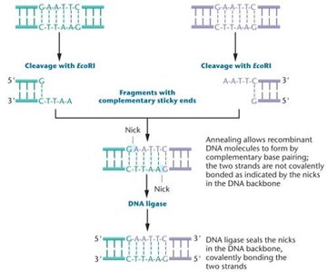
Restriction enzymes are DNA-cutting enzymes produced by bacteria as a defense mechanism against bacteriophage infection. They recognize specific DNA sequences (restriction sites) and cleave both DNA strands, producing restriction fragments. These enzymes are essential for generating DNA fragments with defined ends for cloning.
Palindromic sequences: Recognition sites are often palindromic, meaning the sequence reads the same on both strands in the 5' to 3' direction.
Sticky ends: Overhanging single-stranded ends that can anneal with complementary sequences.
Blunt ends: Double-stranded ends with no overhangs.
DNA Ligase
DNA ligase is an enzyme that covalently joins DNA fragments by sealing the phosphodiester backbone. After restriction enzymes generate compatible ends, DNA ligase is used to create stable recombinant DNA molecules.
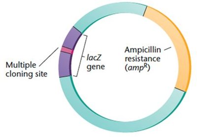
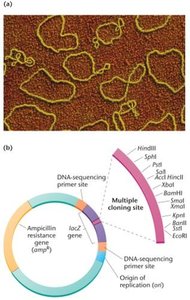
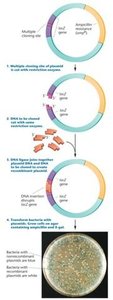
Cloning Vectors
Properties of Cloning Vectors
Cloning vectors are DNA molecules that accept foreign DNA fragments and replicate them in host cells. Essential features include:
Ability to replicate independently of the host chromosome
Multiple restriction enzyme sites (multiple cloning site, MCS)
Selectable marker genes (e.g., antibiotic resistance)
Bacterial Plasmid Vectors
Plasmids are small, circular DNA molecules used as vectors in bacterial cloning. They are engineered to contain MCS, selectable markers, and origins of replication.
Transformation
Transformation is the process of introducing plasmids into bacterial cells. Two main techniques are used:
Calcium ions and heat shock
Electroporation (high-intensity electric pulse)
After transformation, recombinant DNA is replicated within the host cell.
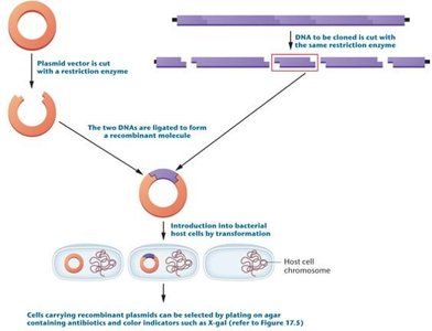
Blue-White Screening
Blue-white screening is a method to distinguish recombinant from nonrecombinant plasmids. Plasmids contain the lacZ gene, which encodes β-galactosidase. When X-gal is present in the medium:
Nonrecombinant plasmids (intact lacZ): blue colonies
Recombinant plasmids (disrupted lacZ): white colonies
Other Types of Cloning Vectors
Phage vectors: Modified bacteriophage genomes, can carry larger DNA fragments (up to 45 kb).
Bacterial Artificial Chromosomes (BACs): Large plasmids, low copy number, can carry 100–300 kb inserts.
Yeast Artificial Chromosomes (YACs): Contain telomeres, centromere, and origin of replication; can carry up to 1000 kb inserts.
Expression Vectors
Expression vectors are designed to ensure transcription and translation of the cloned gene, producing protein in host cells. They are available for both prokaryotic and eukaryotic systems (e.g., Ti plasmid for plants).
Genomic and cDNA Libraries
Genomic Libraries
A genomic library is a collection of DNA fragments representing the entire genome of an organism. It is constructed by cutting genomic DNA with restriction enzymes and cloning the fragments into vectors. Libraries from eukaryotes contain both coding and noncoding sequences.
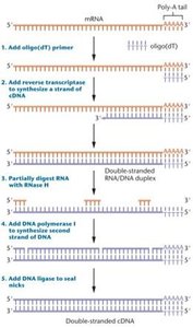
cDNA Libraries
cDNA (complementary DNA) libraries are made from mRNA isolated from cells or tissues. They represent only the expressed genes at the time of isolation and are useful for studying gene expression and identifying genes involved in specific processes (e.g., cancer).
Library Screening
Library screening uses labeled DNA or RNA probes to identify and isolate specific genes of interest from a library. Probes must be complementary to the target sequence and are often tagged for detection.
Polymerase Chain Reaction (PCR)
PCR Principles and Requirements
PCR is a rapid, in vitro method for amplifying specific DNA sequences without the need for host cells. It requires:
Double-stranded target DNA
DNA polymerase (e.g., Taq polymerase)
Primers (short, single-stranded DNA sequences)
Deoxyribonucleoside triphosphates (dNTPs)
Buffer and cofactors (e.g., Mg2+)
PCR Steps
Denaturation: DNA strands are separated by heating.
Annealing: Primers bind to complementary sequences on the single-stranded DNA.
Extension: DNA polymerase synthesizes new DNA strands from the primers.
Each cycle doubles the amount of target DNA, leading to exponential amplification.
Limitations and Applications of PCR
Requires prior knowledge of target sequence for primer design
Highly sensitive to contamination
Cannot efficiently amplify very long DNA segments
Applications include genetic testing, forensics, molecular pathology, and research.
RT-PCR and qPCR
RT-PCR (Reverse Transcription PCR): Used to study gene expression by converting mRNA to cDNA before amplification.
qPCR (Quantitative Real-Time PCR): Allows quantification of DNA amplification in real time.
DNA Analysis Techniques
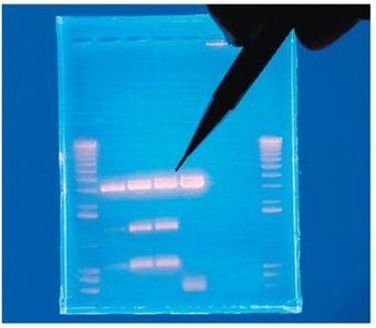
Agarose Gel Electrophoresis
Agarose gel electrophoresis separates DNA fragments by size. DNA is stained (e.g., with ethidium bromide) and visualized under UV light. Smaller fragments migrate farther through the gel.
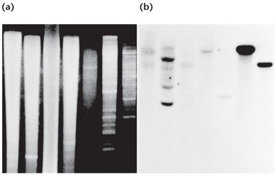
Southern, Northern, and Western Blotting
Southern blot: Detects specific DNA sequences in DNA samples by hybridization with labeled probes after gel electrophoresis and transfer to a membrane.
Northern blot: Used to study RNA expression patterns.
Western blot: Used to detect specific proteins.
Fluorescent in Situ Hybridization (FISH)
FISH uses fluorescently labeled probes to hybridize directly to chromosomes or RNA in cells or tissues, allowing visualization of gene expression or chromosomal location in situ. It is useful in developmental genetics and cytogenetics.
DNA Sequencing Technologies
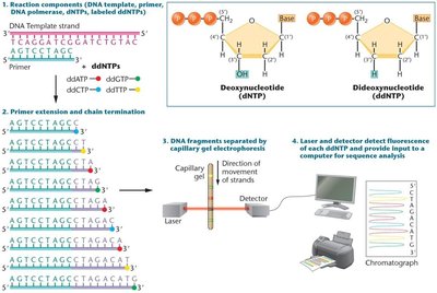
Sanger Sequencing
Sanger sequencing (dideoxynucleotide chain-termination method) is the most common DNA sequencing technique. Incorporation of dideoxynucleotides (ddNTPs) terminates DNA synthesis, generating fragments of varying lengths that can be separated and analyzed to determine the DNA sequence.
Next-Generation and Third-Generation Sequencing
Next-generation sequencing (NGS): Allows simultaneous sequencing of thousands of DNA molecules, generating large datasets rapidly and at lower cost.
Third-generation sequencing (TGS): Sequences single DNA molecules in real time, such as SMRT (Single Molecule Real-Time) sequencing.
Gene Targeting and Genome Editing
Gene Targeting and Knockout Models
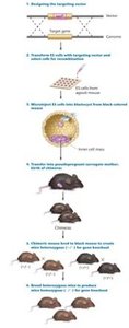
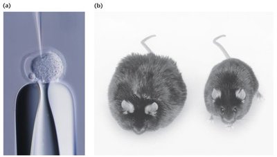
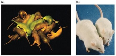
Gene targeting allows specific alleles or sequences to be disrupted or replaced, enabling functional studies. Knockout mice are generated by introducing targeting vectors into embryonic stem (ES) cells, which undergo homologous recombination to disrupt the gene of interest. These ES cells are injected into embryos to produce chimeric and knockout animals.
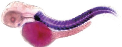
Transgenic Animals
Transgenic (knock-in) animals are engineered to express or overexpress specific genes (transgenes). These models are used to study gene function, regulation, and disease mechanisms.
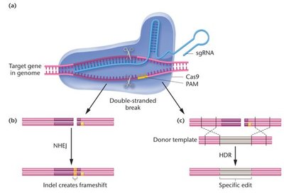
Genome Editing with CRISPR-Cas9
CRISPR-Cas9 is a revolutionary genome editing tool derived from bacterial immune systems. The Cas9 nuclease, guided by a single guide RNA (sgRNA), introduces double-stranded breaks at specific genomic sites adjacent to a PAM sequence (5'-NGG-3'). The cell repairs these breaks via nonhomologous end-joining (NHEJ) or homology-directed repair (HDR), enabling targeted gene disruption or precise editing.
Applications: Gene therapy, crop improvement, disease modeling, and more.
Limitations: Off-target effects, sgRNA specificity, and ongoing improvements in enzyme design.
Summary Table: Key Cloning and Analysis Tools
Tool/Technique | Main Purpose | Key Features |
|---|---|---|
Restriction Enzymes | Cut DNA at specific sequences | Produce sticky/blunt ends |
Cloning Vectors | Carry and replicate DNA fragments | MCS, selectable markers |
PCR | Amplify DNA sequences | Rapid, in vitro, sequence-specific |
Gel Electrophoresis | Separate DNA by size | Visualization under UV |
Southern Blot | Detect specific DNA sequences | Hybridization with probes |
FISH | Visualize gene location/expression | Fluorescent probes |
Sanger Sequencing | Determine DNA sequence | ddNTP chain termination |
CRISPR-Cas9 | Genome editing | Targeted, efficient, versatile |
