 Back
BackChromosomal Abnormalities: Aneuploidy, Structural Variations, and Their Genetic Consequences
Study Guide - Smart Notes
 Tailored notes based on your materials, expanded with key definitions, examples, and context.
Chromosome Number and Structure
Euploidy and Aneuploidy
The number of chromosomes in a nucleus and their structure are species-specific. The euploid number refers to the normal, complete set(s) of chromosomes (e.g., n, 2n, 3n). Aneuploidy describes a chromosome number that is not a complete set, resulting from the addition or loss of one or more chromosomes. These changes can have significant effects on phenotype, development, and viability, especially in animals.
Euploidy: Complete sets of chromosomes (e.g., diploid 2n, triploid 3n).
Aneuploidy: Chromosome number deviates from a complete set (e.g., 2n+1, 2n-1).
Example: Human diploid number is 46; a cell with 47 chromosomes (trisomy) is aneuploid.
Mechanisms of Aneuploidy
Nondisjunction
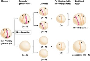
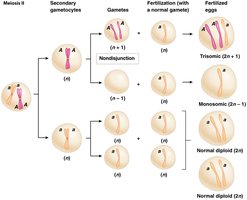
Nondisjunction is the failure of homologous chromosomes or sister chromatids to separate properly during cell division. This can occur in mitosis or meiosis, leading to daughter cells with abnormal chromosome numbers. In meiosis, nondisjunction can occur in either the first or second division, producing gametes that are aneuploid.
Meiosis I Nondisjunction: Failure of homologs to separate; all gametes are abnormal (n+1 or n-1).
Meiosis II Nondisjunction: Failure of sister chromatids to separate; half the gametes are abnormal.
Somatic Nondisjunction: Occurs in mitosis, often seen in cancer cells.
Gene Dosage and Phenotypic Effects
Aneuploidy alters the dosage of all genes on the affected chromosome, leading to phenotypic changes. Most animals are highly sensitive to gene dosage changes, while plants are more tolerant. In humans, only a few autosomal trisomies (chromosomes 13, 18, 21) are compatible with life, and no autosomal monosomies are observed in live births.
Gene Dosage: The number of copies of a gene present in a cell or nucleus.
Trisomy: Three copies of a chromosome (150% gene dosage).
Monosomy: One copy of a chromosome (50% gene dosage).
Human Aneuploidy Syndromes
Trisomy 21 (Down Syndrome)
Down syndrome is caused by trisomy 21 and is associated with intellectual disability and heart defects. The risk increases with maternal age. The Down Syndrome Critical Region (DSCR) on chromosome 21 contains genes such as DYRK and DSCAM that contribute to the phenotype.
Turner Syndrome
Turner syndrome (45, XO) is a monosomy of the X chromosome. The absence of a second sex chromosome leads to developmental abnormalities, primarily due to haploinsufficiency of the SHOX gene.
Other Aneuploidies
Patau syndrome (Trisomy 13): Severe intellectual and physical disabilities.
Edwards syndrome (Trisomy 18): Severe developmental delays and early lethality.
Klinefelter syndrome (47, XXY): Male with extra X chromosome, variable symptoms.
Jacob syndrome (47, XYY): Male with extra Y chromosome, tall stature, minor symptoms.
Triple X syndrome (47, XXX): Female with extra X chromosome, usually mild symptoms.
Fertility and Mosaicism in Aneuploidy
Reduced Fertility in Trisomics
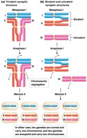
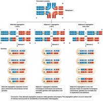
Trisomic individuals often have reduced fertility due to abnormal chromosome segregation during meiosis. Two patterns of synapsis are possible: trivalent and bivalent/univalent arrangements, neither of which segregates chromosomes equally at anaphase I, resulting in semisterility.
Mosaicism
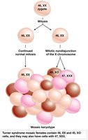
Mosaicism arises from mitotic nondisjunction early in development, resulting in individuals with two or more genetically distinct cell lines. For example, some Turner syndrome individuals are mosaics with both 45, XO and 46, XX cells.
Chromosome Structural Abnormalities
Types of Structural Changes
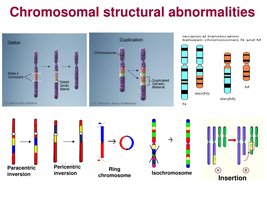
Chromosome breakage can lead to loss, gain, or rearrangement of chromosome segments, causing gene dosage imbalances and potentially severe phenotypic effects. Major types include deletions, duplications, inversions, and translocations.
Deletion: Loss of a chromosome segment.
Duplication: Gain of an extra copy of a chromosome segment.
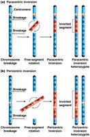
Inversion: Reversal of a chromosome segment (paracentric or pericentric).
Translocation: Segment exchange between nonhomologous chromosomes.
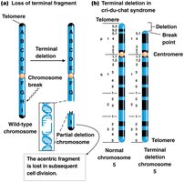
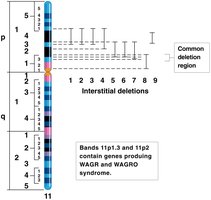
Partial Chromosome Deletions
Deletions can be terminal (loss of an end) or interstitial (loss of an internal segment). Large deletions are detectable microscopically, while smaller ones require molecular techniques for detection. Partial deletion heterozygotes have one normal and one deleted chromosome.
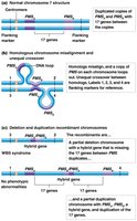
Unequal Crossover
Unequal crossover during meiosis, often due to misalignment of repetitive sequences, can produce partial duplication and deletion heterozygotes. This mechanism is implicated in syndromes such as Williams-Beuren syndrome.
Detection of Duplications and Deletions
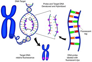
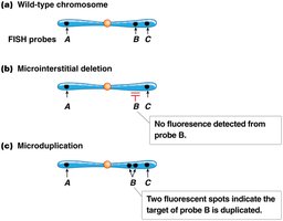
Large chromosomal changes can be seen with microscopy, but microdeletions and microduplications require molecular techniques such as FISH (fluorescent in situ hybridization) for detection.
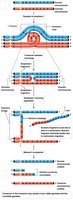
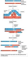
Chromosome Pairing and Recombination in Inversion Heterozygotes
Inversion heterozygotes form an inversion loop during synapsis. Crossing over within the loop can produce dicentric (two centromeres) or acentric (no centromere) chromosomes in paracentric inversions, and chromosomes with duplications and deletions in pericentric inversions. These recombinant chromosomes are usually inviable, leading to crossover suppression.
Chromosome Translocations
Types of Translocations
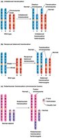
Translocations involve the exchange of chromosome segments between nonhomologous chromosomes. They can be unbalanced, reciprocal balanced, or Robertsonian (chromosome fusion). Translocation heterozygotes may be phenotypically normal but can experience reduced fertility due to abnormal segregation during meiosis.
Unbalanced Translocation: Nonreciprocal transfer of a chromosome segment.
Reciprocal Balanced Translocation: Exchange of segments between two nonhomologous chromosomes.
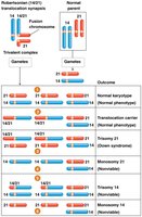
Robertsonian Translocation: Fusion of two acrocentric chromosomes, reducing chromosome number.
Segregation Patterns in Translocation Heterozygotes
During meiosis, translocation heterozygotes form a cross-shaped structure. Alternate segregation produces viable gametes, while adjacent segregation leads to gametes with duplications and deletions, causing semisterility.
Robertsonian Translocation and Familial Down Syndrome
Robertsonian translocation between chromosomes 21 and 14 can result in familial Down syndrome. Carriers are usually phenotypically normal but have an increased risk of producing offspring with Down syndrome due to abnormal segregation.
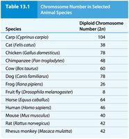
Summary Table: Chromosome Number in Selected Animal Species
Species | Diploid Chromosome Number (2n) |
|---|---|
Carp (Cyprinus carpio) | 104 |
Cat (Felis catus) | 38 |
Chicken (Gallus domesticus) | 78 |
Chimpanzee (Pan troglodytes) | 48 |
Cow (Bos taurus) | 60 |
Dog (Canis familiaris) | 78 |
Frog (Rana pipiens) | 26 |
Fruit fly (Drosophila melanogaster) | 8 |
Horse (Equus caballus) | 64 |
Human (Homo sapiens) | 46 |
Mouse (Mus musculus) | 40 |
Rat (Rattus norvegicus) | 42 |
Rhesus monkey (Macaca mulatta) | 42 |
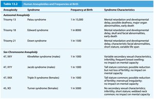
Summary Table: Human Aneuploidies and Frequencies at Birth
Aneuploidy | Syndrome | Frequency at Birth | Syndrome Characteristics |
|---|---|---|---|
Trisomy 13 | Patau syndrome | 1 in 15,000 | Mental retardation, developmental delay, polydactyly, cleft palate, organ defects, short life span |
Trisomy 18 | Edwards syndrome | 1 in 8,000 | Mental retardation, developmental delay, skull and facial abnormalities, early lethality |
Trisomy 21 | Down syndrome | 1 in 800 | Mental retardation, developmental delay, characteristic facial features, heart defects, variable life span |
47, XXY | Klinefelter syndrome (males) | 1 in 1,000 | Variable secondary sexual characteristics, infertility, tall stature, mild cognitive effects |
47, XYY | Jacob syndrome (males) | 1 in 1,000 | Tall stature, common, possible reduction in fertility, minor cognitive effects |
47, XXX | Triple X syndrome (females) | 1 in 1,000 | Tall stature, common, possible reduction in fertility, minor cognitive effects |
45, XO | Turner syndrome (females) | 1 in 5,000 | No secondary sexual characteristics, infertility, short stature, normal intelligence |
