 Back
Chromosomal Organization of DNA: Structure and Function in Prokaryotes and Eukaryotes
Study Guide - Smart Notes
 Tailored notes based on your materials, expanded with key definitions, examples, and context.
Chromosomal Organization of DNA
Viral and Prokaryotic Chromosomes
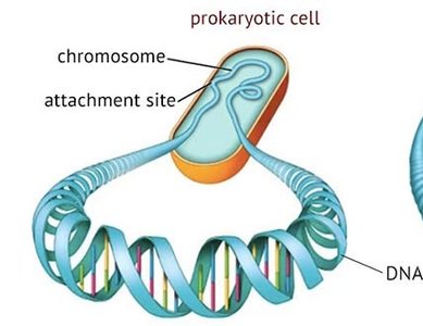
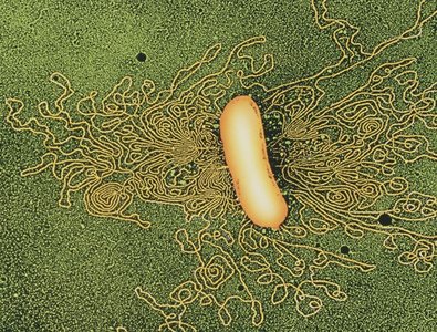
Viral and prokaryotic chromosomes are generally composed of a single molecule of nucleic acid, either DNA or RNA in viruses, and DNA in prokaryotes. Their chromosomal structure is much simpler than that of eukaryotes, with fewer associated proteins and less genetic information.
Viral nucleic acids: Can be single-stranded (ss) or double-stranded (ds), and may be circular or linear.
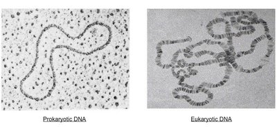
Prokaryotic DNA: Typically double-stranded, circular, and compacted into the nucleoid region of the cell.
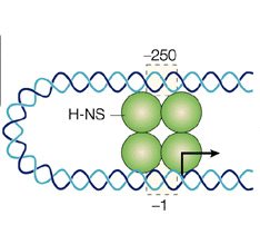
Associated proteins: Proteins such as H-NS and HU help compact prokaryotic DNA, similar to histones in eukaryotes.
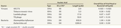
Comparison Table: Viral and Bacterial Nucleic Acids
Source | Nucleic Acid Type | SS or DS | Length (µm) | Overall Size (µm) |
|---|---|---|---|---|
φX174 | DNA | SS | 2.0 | 0.025 x 0.025 |
Tobacco mosaic virus | RNA | SS | 3.3 | 0.03 x 0.02 |
Lambda phage | DNA | DS | 17.0 | 0.067 x 0.10 |
T2 phage | DNA | DS | 52.0 | 0.10 x 0.10 |
Haemophilus influenzae | DNA | DS | 820.0 | 1.00 x 0.30 |
Escherichia coli | DNA | DS | 1200.0 | 2.00 x 0.50 |
Supercoiling and Topoisomers
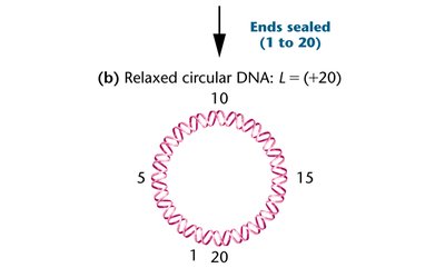
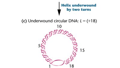
Supercoiling is a process that compacts DNA further, and can occur in both viral and bacterial chromosomes. Molecules with different degrees of coiling are called topoisomers. The enzyme topoisomerase is responsible for introducing or removing supercoils.
Relaxed circular DNA: Has a specific linking number (L).
Underwound DNA: Has fewer helical turns, increasing the potential for supercoiling.
Specialized Chromosomes: Polytene and Lampbrush Chromosomes
Polytene Chromosomes
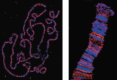
Polytene chromosomes are very large chromosomal structures found in certain tissues, such as the salivary glands of Drosophila. They are visible during interphase and are characterized by distinctive banding patterns.
Structure: Made of paired homologs and many identical DNA strands (1000–5000), resulting from multiple rounds of replication without cell division.
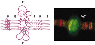
Banding: Alternating bands (chromomeres) are visible under the light microscope.
Puffs: Areas of uncoiling, called puffs, indicate regions of active transcription.
Lampbrush Chromosomes
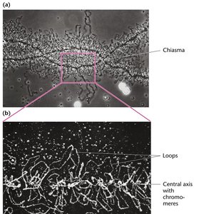
Lampbrush chromosomes are elongated meiotic chromosomes found in oocytes of vertebrates and some insects. They are characterized by extensive looping, which increases the surface area for transcription.
Structure: Made of two sister chromatids with loops extending from a central axis.
Function: Direct metabolism and increase gene expression during oogenesis.
Eukaryotic Chromosomes and Chromatin Structure
Chromatin Organization
During interphase, eukaryotic chromosomes decondense into chromatin, a complex of DNA, RNA, histone proteins, and non-histone proteins. Chromatin is distributed throughout the nucleus and is much longer when decondensed.
Chromatin: The mixture of DNA and proteins that forms chromosomes in eukaryotic cells.
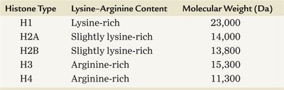
Histones: Positively charged proteins rich in lysine and arginine, essential for chromatin structure.
Non-histone proteins: Contribute to chromatin structure and function.
Histone Types and Properties
Histone Type | Lysine–Arginine Content | Molecular Weight (Da) |
|---|---|---|
H1 | Lysine-rich | 23,000 |
H2A | Slightly lysine-rich | 14,000 |
H2B | Slightly lysine-rich | 13,800 |
H3 | Arginine-rich | 15,300 |
H4 | Arginine-rich | 11,300 |
Nucleosome Structure
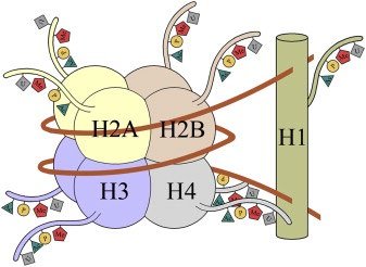
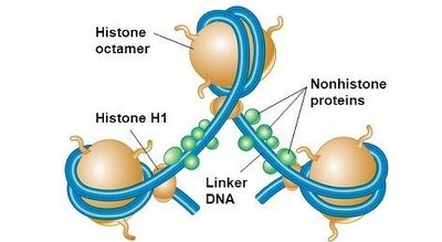
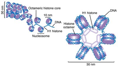
The basic unit of chromatin is the nucleosome, consisting of DNA wrapped around a histone octamer. Nucleosomes are connected by linker DNA and further compacted into higher-order structures.
Nucleosome: Contains two copies each of H2A, H2B, H3, and H4, with about 200 base pairs of DNA.
Histone H1: Associated with linker DNA between nucleosomes.
Chromatin fiber: Six nucleosomes form a 30 nm fiber, which coils further into a 300 nm chromatid.
Chromatin Remodeling and Transcription
Chromatin Remodeling
Chromatin remodeling is the process by which chromatin structure is altered to allow access to DNA for transcription. This is essential for gene expression, as tightly wound DNA is inaccessible to transcription machinery.
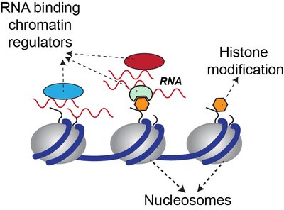
Histone tails: Can be chemically modified, affecting chromatin structure and gene expression.
Remodeling proteins: Interact with nucleosomes to reposition or remove histones.
Chemical Modifications of Histones
Histone proteins can undergo several chemical modifications, which influence chromatin structure and transcriptional activity.
Acetylation: Addition of acetyl groups to lysine residues by histone acetyltransferase (HAT), neutralizing positive charge and loosening DNA-histone interaction.
Methylation: Addition of methyl groups to lysine or arginine by methyltransferase, which can either activate or repress transcription depending on context.
Phosphorylation: Addition of phosphate groups to serine or histidine by kinases, introducing negative charge and affecting chromatin structure.
Euchromatin vs. Heterochromatin
Euchromatin
Euchromatin is the relatively uncoiled form of chromatin, which is accessible for transcription and contains actively expressed genes.
Heterochromatin
Heterochromatin is tightly coiled chromatin, generally unavailable for expression. It tends to lack genes or contain unexpressed genes and includes regions such as telomeres and centromeres.
Barr body: In human females, one X chromosome is condensed into a Barr body, a form of heterochromatin.
Y chromosome: Most of the Y chromosome in human males is inert heterochromatin.
Summary Table: Prokaryotic vs. Eukaryotic DNA Organization
Feature | Prokaryotes | Eukaryotes |
|---|---|---|
Chromosome Structure | Single, circular DNA | Multiple, linear chromosomes |
Associated Proteins | Few (H-NS, HU) | Many (histones, non-histone proteins) |
DNA Compaction | Nucleoid region | Chromatin, nucleosomes |
Cell Division | No mitosis | Mitosis and meiosis |
Gene Expression | Less regulation | Complex regulation |
Key Equations
Linking Number (L): Where T is the number of helical turns and W is the number of supercoils.
DNA Fragment Protection: Endonuclease digestion yields fragments of approximately 200 base pairs, indicating nucleosome protection.
Conclusion
The organization of DNA within chromosomes varies greatly between prokaryotes, viruses, and eukaryotes. Eukaryotic chromatin structure is highly complex, involving nucleosomes, histones, and various levels of compaction. Chromatin remodeling and histone modifications are essential for regulating gene expression, and specialized chromosomes such as polytene and lampbrush chromosomes provide insight into chromosomal organization and function.
