 Back
Chromosomal Organization of DNA: Structure and Function in Prokaryotes, Viruses, and Eukaryotes
Study Guide - Smart Notes
 Tailored notes based on your materials, expanded with key definitions, examples, and context.
Chromosomal Organization of DNA
Viral and Prokaryotic Chromosomes
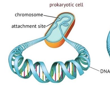
The structure of DNA in non-eukaryotic cells, such as viruses and prokaryotes, is relatively simple compared to eukaryotes. These chromosomes typically consist of a single molecule of nucleic acid, either DNA or RNA in viruses, and DNA in prokaryotes. The amount of genetic information is much less than in eukaryotic cells, and there are fewer associated proteins.
Viral nucleic acids: Can be single-stranded (ss) or double-stranded (ds), and may be circular or linear.
Prokaryotic DNA: Usually double-stranded, compacted into a nucleoid region, and associated with proteins such as H-NS and HU.
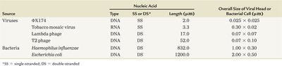
Comparison Table: Viral and Bacterial Nucleic Acids
Source | Nucleic Acid Type | SS or DS | Length (µm) | Overall Size (µm) |
|---|---|---|---|---|
φX174 | DNA | SS | 2.0 | 0.025 x 0.025 |
Tobacco mosaic virus | RNA | SS | 3.3 | 0.03 x 0.02 |
Lambda phage | DNA | DS | 17.0 | 0.07 x 0.07 |
T2 phage | DNA | DS | 52.0 | 0.067 x 0.110 |
Haemophilus influenzae | DNA | DS | 820.0 | 1.00 x 0.30 |
Escherichia coli | DNA | DS | 1200.0 | 2.00 x 0.50 |
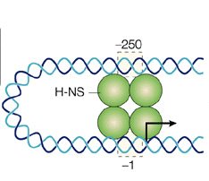
Bacterial DNA Packaging and DNA-Binding Proteins
Bacterial DNA is compacted and structured within the nucleoid region. Proteins such as H-NS and HU, which are similar to eukaryotic histones, play a crucial role in folding and compacting DNA. These proteins have positively charged amino acid residues that attract the negatively charged phosphate groups of DNA, facilitating compaction.
H-NS and HU: Histone-like proteins that help organize and compact bacterial DNA.
Compaction: Essential for fitting large DNA molecules into small cellular spaces.
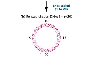
Supercoiling and Topoisomerases
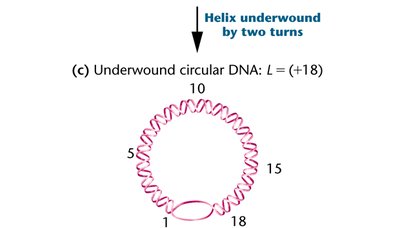
Supercoiling is a process that further compacts DNA in both viral and bacterial chromosomes. Identical DNA molecules with different degrees of coiling are called topoisomers. The enzyme responsible for introducing or removing supercoils is topoisomerase.
Relaxed circular DNA: No supercoiling, normal helical turns.
Underwound DNA: Fewer helical turns, introduces negative supercoiling.
Topoisomerase: Enzyme that regulates DNA supercoiling.
Specialized Chromosomes: Polytene and Lampbrush Chromosomes
Polytene Chromosomes
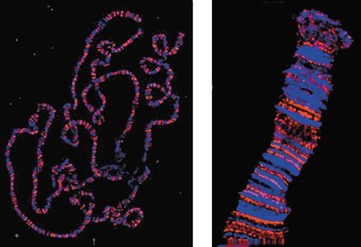
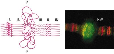
Polytene chromosomes are very large chromosomal structures that can be observed during interphase using a light microscope. They are made of paired homologs and consist of many identical DNA strands, resulting from multiple rounds of replication without strand separation or cytoplasmic division. These chromosomes display distinctive linear alternating bands, known as chromomeres, and areas of uncoiling called puffs, which are sites of active transcription.
Banding pattern: Caused by parallel arrangement of DNA strands.
Puffs: Regions of uncoiling associated with increased gene expression.
Lampbrush Chromosomes
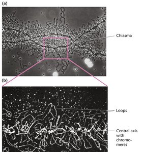
Lampbrush chromosomes are another form of specialized chromosome, initially discovered in amphibian oocytes but now known to be common in vertebrate oocytes and some insect spermatocytes. These are meiotic chromosomes that elongate during use, facilitating direct metabolism and increased gene expression. They are composed of two sister chromatids and display loops that are sites of active transcription.
Loops: Extend from the central axis, representing regions of active transcription.
Chromomeres: Linear arrangement along the central axis.
Comparison of Prokaryotic/Viral and Eukaryotic DNA
Eukaryotic Chromosomes and Chromatin Structure
Eukaryotic chromosomes are much more complex than prokaryotic or viral chromosomes. During interphase, chromosomes decondense into chromatin, a mixture of DNA, RNA, histone proteins, and non-histone proteins. Chromatin is distributed throughout the nucleus and is approximately 10,000 times longer than when condensed. Eukaryotic DNA is associated with a large number of proteins, including histones, which are essential for chromatin structure.
Histones: Positively charged proteins rich in lysine and arginine, crucial for DNA packaging.
Chromatin: The complex of DNA and proteins found in eukaryotic cells.
Chromosome length: Human chromosomes can be stretched to ~2 meters, yet all fit within a 5–10 µm nucleus.
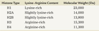
Histone Types and Properties
Histone Type | Lysine–Arginine Content | Molecular Weight (Da) |
|---|---|---|
H1 | Lysine-rich | 23,000 |
H2A | Slightly lysine-rich | 14,000 |
H2B | Slightly lysine-rich | 13,800 |
H3 | Arginine-rich | 15,300 |
H4 | Arginine-rich | 11,300 |
Nucleosome Structure and Chromatin Organization
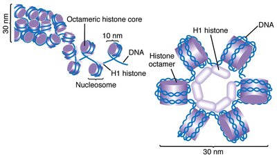
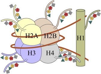
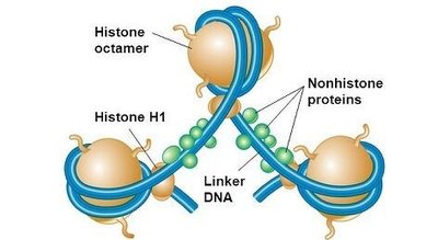
Nucleosomes are the fundamental units of chromatin, consisting of DNA wrapped around histone octamers. Each nucleosome contains two copies each of H2A, H2B, H3, and H4, with approximately 200 base pairs of DNA. Further digestion reveals 147 bp fragments, indicating that linker DNA connects nucleosomes and is associated with histone H1. Chromatin fibers are organized hierarchically, from nucleosomes to 30 nm fibers, then to 300 nm fibers, and finally to chromatids.
Nucleosome: DNA wrapped around histone octamer.
Linker DNA: Connects nucleosomes, associated with H1 histone.
Chromatin fiber: Higher-order structure formed by coiling nucleosomes.
Chromatin Remodeling and Transcription
Chromatin Remodeling
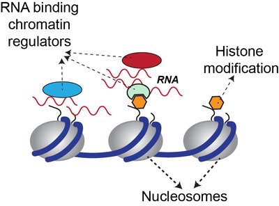
Transcription requires that DNA be accessible to proteins involved in gene expression. Chromatin remodeling is the process by which chromatin structure is altered to allow transcription. This involves direct interaction of proteins with DNA and chemical modifications of histone tails, such as acetylation, methylation, and phosphorylation.
Acetylation: Addition of acetyl groups to lysine, neutralizing positive charge and loosening DNA-histone interaction.
Methylation: Addition of methyl groups to lysine or arginine, affecting chromatin structure and gene expression.
Phosphorylation: Addition of phosphate groups to serine or histidine, introducing negative charge.
Euchromatin vs. Heterochromatin
Chromatin States
Chromatin exists in two main states: euchromatin and heterochromatin. Euchromatin is relatively uncoiled and available for gene expression, while heterochromatin is tightly coiled and generally inactive. Heterochromatin includes regions such as telomeres and centromeres, and is important for structural integrity and gene regulation.
Euchromatin: Active, uncoiled, gene-rich regions.
Heterochromatin: Inactive, tightly coiled, gene-poor regions.
Barr body: Inactive X chromosome in female mammals, condensed as heterochromatin.
Summary Table: Key Differences Between Prokaryotic and Eukaryotic DNA
Feature | Prokaryotic DNA | Eukaryotic DNA |
|---|---|---|
Structure | Single, circular molecule | Multiple, linear chromosomes |
Associated Proteins | Few, histone-like | Many, histones and non-histones |
Compaction | Nucleoid region | Chromatin, nucleosomes |
Gene Expression | Direct, less regulated | Complex, regulated by chromatin state |
Additional info: Chromatin remodeling is a dynamic process essential for gene regulation, development, and cellular differentiation. Disruption of chromatin structure can lead to diseases such as cancer.
