 Back
BackChromosomal Structure, Variation, and Experimental Methods in Human Genetics
Study Guide - Smart Notes
 Tailored notes based on your materials, expanded with key definitions, examples, and context.
שינויים במספר ובמבנה הכרומוזומים (Chromosomal Number and Structural Variations)
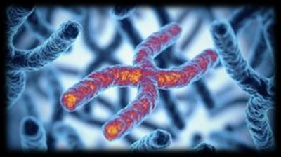
מבנה הכרומוזום (Chromosome Structure)
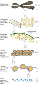
Chromosomes are highly condensed structures of DNA and proteins, essential for the storage, transmission, and regulation of genetic information. Their organization allows for efficient segregation during cell division and protection of genetic material.
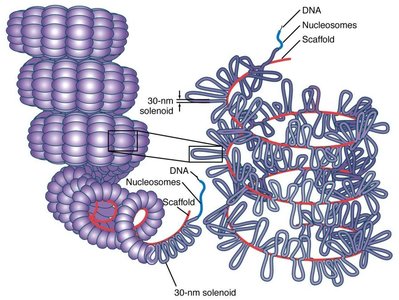
DNA Packaging: DNA is wrapped around histone proteins, forming nucleosomes, which further coil and fold to create the compact chromosome structure.
Key Elements: Each chromosome contains a centromere, telomeres at both ends, and multiple origins of replication (ORIs).
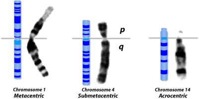
Chromosome Arms: The centromere divides the chromosome into a short arm (p) and a long arm (q).
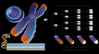
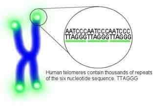
טלומרים (Telomeres)
Telomeres are repetitive nucleotide sequences at the ends of chromosomes, protecting them from degradation and fusion with other chromosomes. In humans, the repeat sequence is TTAGGG, spanning 3-20 kb.
Function: Maintain chromosomal integrity and prevent end-to-end fusions.
Telomerase: An enzyme that extends telomeres, counteracting their shortening during cell division.
Cellular Aging: Progressive telomere shortening leads to cellular senescence or apoptosis.
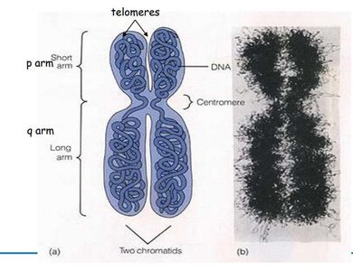
צנטרומר (Centromere)
The centromere is a specialized chromosomal region composed of repetitive DNA sequences. It is essential for the correct segregation of chromosomes during cell division, serving as the attachment site for spindle fibers.
Location: Divides the chromosome into p (short) and q (long) arms.
Function: Ensures proper movement of chromosomes during mitosis and meiosis.

סיווג כרומוזומים (Chromosome Classification)
Human chromosomes are classified based on length, centromere position, and banding pattern:
Metacentric: Centromere in the middle, arms of equal length.
Submetacentric: Centromere slightly off-center, creating unequal arms.
Acrocentric: Centromere near one end, resulting in a very short p arm.
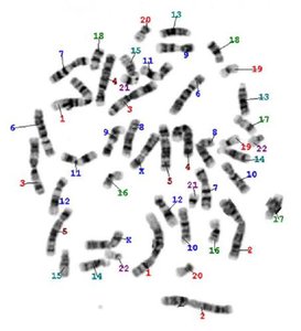
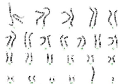
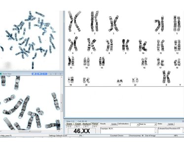
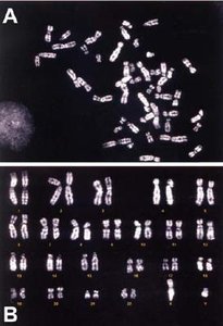
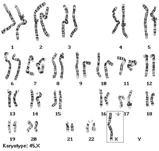
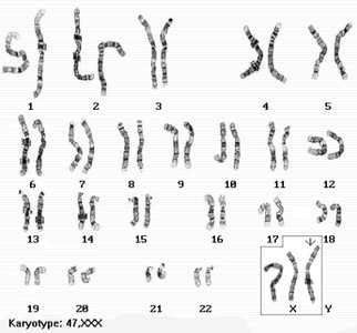
ציטוגנטיקה וקריוטיפ (Cytogenetics and Karyotyping)
הגדרות בסיסיות (Basic Definitions)
ציטוגנטיקה (Cytogenetics): The microscopic study of chromosomes.
קריוטיפ (Karyotype): The complete set of chromosomes in a cell, organized by size and morphology.
אידיוגרמה (Idiogram): A schematic representation of the karyotype, showing banding patterns and chromosome structure.
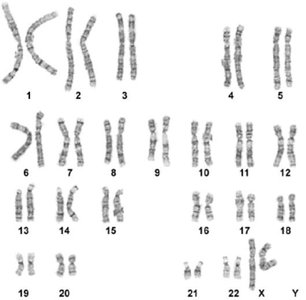
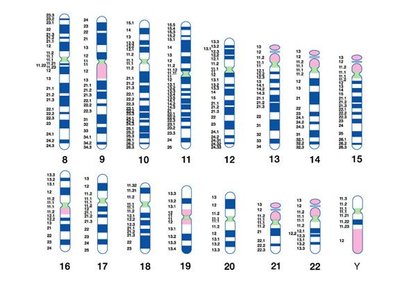
צביעת כרומוזומים (Chromosome Banding Techniques)
Chromosome banding techniques are used to visualize structural features and identify chromosomal abnormalities.
Giemsa (G) Banding: Most common; produces characteristic dark and light bands. Dark bands are gene-poor, light bands are gene-rich.
Q (Quinacrine) Banding: Fluorescent staining, useful for Y chromosome and highly condensed regions.
R (Reverse) Banding: Highlights GC-rich regions, useful for X chromosome analysis.
מבנה הכרומטין (Chromatin Structure)
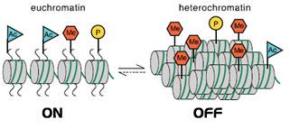
אאוכרומטין והטרוכרומטין (Euchromatin and Heterochromatin)
Chromatin exists in two main forms:
אאוכרומטין (Euchromatin): Less condensed, transcriptionally active, stains lightly.
הטרוכרומטין (Heterochromatin): Highly condensed, transcriptionally inactive, stains darkly. Includes constitutive (always condensed, e.g., centromeres, telomeres) and facultative (can switch states, e.g., inactive X chromosome in females).
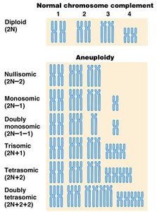
שינויים במספר הכרומוזומים (Chromosomal Number Variations)
אנאופלואידיה (Aneuploidy)
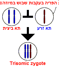
Aneuploidy refers to the presence of an abnormal number of chromosomes in a cell, often resulting from nondisjunction during meiosis.
Nullisomy (2n-2): Loss of both homologs of a chromosome pair (lethal).
Monosomy (2n-1): Loss of a single chromosome (usually lethal except for X chromosome).
Trisomy (2n+1): One extra chromosome (e.g., Down syndrome).
Tetrasomy (2n+2): Two extra homologous chromosomes.
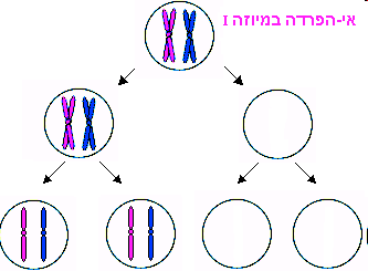
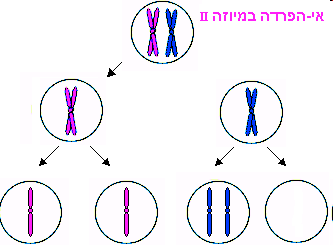
אי-הפרדה (Nondisjunction)
Nondisjunction is the failure of homologous chromosomes or sister chromatids to separate properly during meiosis, leading to gametes with abnormal chromosome numbers.
Meiosis I Nondisjunction: Both homologs go to one gamete; results in two gametes with an extra chromosome and two with none.
Meiosis II Nondisjunction: Sister chromatids fail to separate; results in two normal gametes, one with an extra chromosome, and one missing a chromosome.
דוגמאות לאנאופלואידיות באדם (Examples of Human Aneuploidies)
Turner Syndrome (45,X): Monosomy X; phenotypically female, short stature, infertility.
Triple X Syndrome (47,XXX): Extra X chromosome in females; usually normal phenotype.
Klinefelter Syndrome (47,XXY): Extra X chromosome in males; tall stature, infertility.
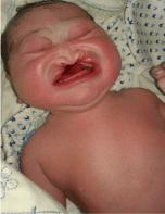
Down Syndrome (Trisomy 21): Most common viable autosomal trisomy; intellectual disability, characteristic facial features, increased risk of congenital heart defects and leukemia.
Edwards Syndrome (Trisomy 18): Severe developmental delays, organ defects, low survival rate.
Patau Syndrome (Trisomy 13): Severe intellectual disability, cleft lip/palate, polydactyly, low survival rate.
שינויים במבנה הכרומוזום (Chromosomal Structural Variations)
סוגי אברציות מבניות (Types of Structural Aberrations)
Structural changes in chromosomes arise from breakage and incorrect rejoining of DNA segments, often during meiosis. These include:
Deletion: Loss of a chromosome segment (e.g., Cri du Chat syndrome).
Duplication: Repetition of a chromosome segment.
Inversion: A chromosome segment is reversed end to end.
Translocation: A segment from one chromosome is transferred to another (e.g., Philadelphia chromosome in chronic myeloid leukemia).

שיטות ניסיוניות בגנטיקה (Experimental Methods in Genetics)
בדיקת קריוטיפ (Karyotyping)
Karyotyping involves the microscopic examination of chromosomes to detect numerical and structural abnormalities. It is a standard method in prenatal diagnosis and cancer genetics.
שיטת FISH (Fluorescence In Situ Hybridization)
FISH uses fluorescent probes to detect specific DNA sequences on chromosomes, allowing for the identification of submicroscopic deletions, duplications, and rearrangements.
Applications: Detection of microdeletions (e.g., DiGeorge syndrome), gene mapping, and cancer diagnostics.
צ'יפ גנטי (Genetic Microarray)
Microarrays can detect copy number variations (duplications and deletions) across the genome with high resolution, identifying subtle genetic changes not visible by karyotyping.
אלקטרופורזה בג'ל (Gel Electrophoresis)
Gel electrophoresis separates DNA fragments by size using an electric field. Smaller fragments migrate faster through the gel matrix.
Applications: DNA fingerprinting, mutation detection, and restriction fragment length polymorphism (RFLP) analysis.
RFLP (Restriction Fragment Length Polymorphism)
RFLP analysis uses restriction enzymes to cut DNA at specific sequences. Variations in restriction sites among individuals create different fragment patterns, useful for genetic mapping and disease diagnosis.
ריצוף DNA (DNA Sequencing)
Modern sequencing technologies allow for the complete determination of the nucleotide sequence of DNA, enabling the detection of point mutations, insertions, deletions, and mosaicism.
טבלת סיווג אנאופלואידיות (Aneuploidy Classification Table)
Type | Chromosome Number | Description |
|---|---|---|
Diploid | 2n | Normal chromosome complement |
Nullisomic | 2n-2 | Loss of both homologs of a chromosome pair |
Monosomic | 2n-1 | Loss of a single chromosome |
Trisomic | 2n+1 | One extra chromosome |
Tetrasomic | 2n+2 | Two extra homologous chromosomes |
סיכום (Summary)
Understanding chromosomal structure, number, and experimental analysis is fundamental to genetics. Chromosomal abnormalities, both numerical and structural, are major contributors to genetic diseases and developmental disorders. Modern cytogenetic and molecular techniques enable precise diagnosis and research into the mechanisms underlying these variations.
