 Back
BackChromosome Number, Structure, and Abnormalities in Eukaryotes
Study Guide - Smart Notes
 Tailored notes based on your materials, expanded with key definitions, examples, and context.
Chromosome Number and Shape in Eukaryotes
Variation in Chromosome Number Among Species
Chromosome number and morphology vary widely among eukaryotic organisms. The diploid chromosome number (2n) is species-specific and is a fundamental genetic characteristic.
Diploid Number (2n): The total number of chromosomes in a somatic cell, representing two sets—one from each parent.
Examples: Humans (2n = 46), Fruit fly (2n = 8), Carp (2n = 104).
Chromosome number does not correlate with organismal complexity.
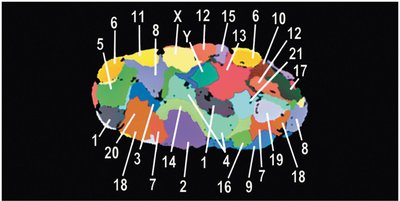
Chromosome Territories in the Nucleus
Chromosomes are not randomly distributed within the nucleus. Instead, each chromosome occupies a distinct region called a chromosome territory during interphase.
Chromosome territories are stable during interphase but change during mitosis.
Chromosomes are anchored by their centromeres and are dynamic within their territories, moving during transcription and DNA replication.
Interchromosomal domains are channels between territories, facilitating the movement of proteins, enzymes, and RNA molecules.
Larger, gene-rich chromosomes are typically located near the nuclear center, while smaller, gene-poor chromosomes are near the periphery.
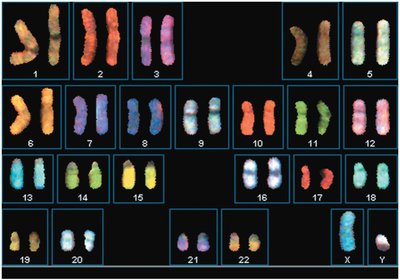
Chromosome Visualization and Karyotypes
Chromosome condensation peaks at metaphase, allowing for visualization and identification using microscopy and molecular techniques.
Karyotype: An organized visual display of chromosomes, arranged by size and shape, used to detect abnormalities in number or structure.
Homologous chromosomes are paired and ordered by descending size.
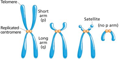
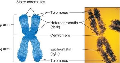
Chromosome Shape and Centromere Position
Chromosome shape is determined by the position of the centromere, which divides the chromosome into two arms:
p arm: Short arm
q arm: Long arm
Types of chromosomes based on centromere position:
Metacentric: Centromere near the middle
Submetacentric: Centromere between center and tip
Acrocentric: Centromere close to one end
Telocentric: Centromere at the tip (no p arm)
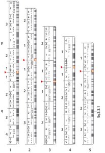
Chromosome Banding and Identification
Chromosome banding techniques allow cytogeneticists to distinguish chromosomes based on size, shape, and banding pattern.
Giemsa (G) banding: The standard for human chromosomes, producing distinct, reproducible patterns.
Banding patterns are used to identify structural abnormalities.
Heterochromatin and Euchromatin
Chromosome condensation varies along its length, affecting gene expression:
Euchromatin: Less condensed, gene-rich, and actively transcribed regions.
Heterochromatin: Highly condensed, gene-poor, and transcriptionally inactive regions.
Polyploidy and Changes in Chromosome Number
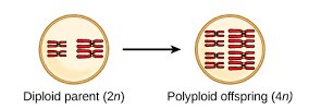
Polyploidy: Definition and Types
Polyploidy is the presence of three or more complete sets of chromosomes in the nucleus. It is common in plants and can arise by different mechanisms:
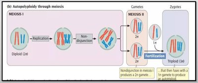
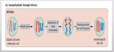
Autopolyploidy: Duplication of chromosome sets within a single species.
Allopolyploidy: Combination of chromosome sets from different species.
Mechanisms of Polyploidy Formation
Meiotic Nondisjunction: Failure of homologous chromosomes to separate, producing diploid gametes. Fusion with a normal gamete yields triploid or tetraploid offspring.
Mitotic Nondisjunction: Failure of sister chromatids to separate during mitosis, doubling chromosome number in somatic cells.
Consequences and Applications of Polyploidy
Polyploid plants often have larger fruits and flowers.
Odd-numbered polyploids (e.g., 3n) are usually sterile, useful for producing seedless varieties.
Polyploidy can lead to rapid speciation and is a major evolutionary force in plants.
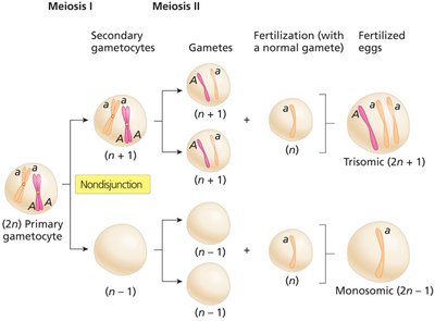
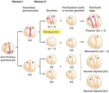
Nondisjunction and Aneuploidy
Nondisjunction: Definition and Effects
Nondisjunction is the failure of chromosomes or sister chromatids to separate properly during cell division, leading to abnormal chromosome numbers (aneuploidy).
Aneuploidy: Presence of an abnormal number of chromosomes (not a complete set).
Common forms: Monosomy (2n-1) and Trisomy (2n+1).
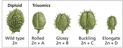
Gene Dosage and Phenotypic Effects
Changes in gene dosage due to aneuploidy can disrupt the balance of gene products, often resulting in severe phenotypic consequences, especially in animals.
Plants tolerate gene dosage changes better than animals.
Example: Trisomic Datura plants show distinct phenotypes for each trisomy.
Aneuploidy in Humans
Most human aneuploidies are lethal, but some survive to birth with characteristic syndromes:
Aneuploidy | Syndrome | Frequency at Birth | Characteristics |
|---|---|---|---|
Trisomy 21 | Down syndrome | 1 in 1500 | Mental retardation, facial abnormalities, short stature |
Trisomy 18 | Edwards syndrome | 1 in 8000 | Developmental delay, organ abnormalities |
Trisomy 13 | Patau syndrome | 1 in 15,000 | Developmental delay, organ abnormalities |
47, XXY | Klinefelter syndrome | 1 in 1000 | Male infertility, breast swelling |
47, XYY | Jacob syndrome | 1 in 1000 | Tall stature, possible fertility reduction |
47, XXX | Triple X syndrome | 1 in 1000 | Tall stature, possible fertility reduction |
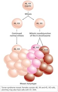
45, XO | Turner syndrome | 1 in 5000 | Female infertility, short stature, webbed neck |
Special Cases: Mosaicism and Uniparental Disomy
Mosaicism: Presence of two or more genetically distinct cell lines in an individual, often due to mitotic nondisjunction.
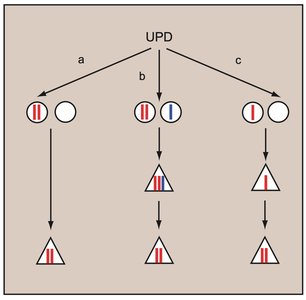
Uniparental Disomy: Both copies of a chromosome are inherited from one parent, associated with disorders like Prader-Willi and Angelman syndromes.
Chromosome Structural Mutations
Chromosome Breakage and Rearrangement
Breakage of chromosomes can lead to loss, gain, or rearrangement of genetic material, causing gene dosage imbalances and potentially severe phenotypic effects.
Deletions: Loss of a chromosome segment; can be terminal (end) or interstitial (internal).
Duplications: Gain of a chromosome segment, often due to unequal crossover.
Inversions: Reversal of a chromosome segment; can be paracentric (excluding centromere) or pericentric (including centromere).
Translocations: Movement of a chromosome segment to a nonhomologous chromosome; can be reciprocal or Robertsonian.
Detection and Genetic Implications
Large deletions/duplications can be detected by altered banding patterns; microdeletions require molecular techniques like FISH.
Inversion and translocation heterozygotes may have reduced fertility due to abnormal meiotic segregation.
Position effect variegation demonstrates that gene expression can be influenced by chromatin structure and location.
Chromatin Organization in Eukaryotic Chromosomes
Chromatin Structure and Compaction
Eukaryotic chromosomes are composed of DNA and proteins, organized into chromatin. Chromatin compaction is essential for chromosome function, segregation, and gene regulation.
Histones: Small, basic proteins (H1, H2A, H2B, H3, H4) that form the core of nucleosomes.
Nucleosome: Fundamental unit of chromatin, consisting of DNA wrapped around a histone octamer.
Chromatin fibers further coil into higher-order structures (30-nm fiber, chromatin loops, metaphase chromosome).
Role of Chromatin in Gene Expression
Chromatin state (euchromatin vs. heterochromatin) regulates access to DNA for transcription.
Position effect variegation in Drosophila illustrates how gene expression is silenced when a gene is relocated near heterochromatin.
Chromatin structure is heritable and can influence gene expression across cell generations.
