 Back
BackChromosome Structure and DNA Organization: Study Notes
Study Guide - Smart Notes
 Tailored notes based on your materials, expanded with key definitions, examples, and context.
Chromosome Structure and DNA Organization
Introduction to Chromosome Structure
The organization of DNA within cells is fundamental to genetic function and inheritance. DNA is packaged into genes, which are further organized into chromosomes. Advances in microscopy have enabled detailed analysis of chromosome structure in viruses, bacteria, and eukaryotes, including specialized forms such as polytene and lampbrush chromosomes.
Viral and Bacterial Chromosomes
Structural Organization
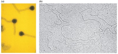
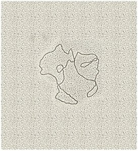
Viral chromosomes consist of a single nucleic acid molecule, which may be DNA or RNA, and can be single- or double-stranded, circular or linear.
Bacterial chromosomes are typically circular, double-stranded DNA molecules compacted into a region called the nucleoid.
Both viral and bacterial chromosomes are largely devoid of associated proteins, making them structurally simpler than eukaryotic chromosomes.
These chromosomes are much smaller and contain less genetic information compared to eukaryotic chromosomes.
Examples of Viral and Bacterial Genetic Material
Organism | Nucleic Acid Type | SS or DS* | Nucleic Acid Length (µm) | Overall Size (µm) |
|---|---|---|---|---|
ΦX174 (virus) | DNA | SS | 2.0 | 0.025 × 0.025 |
Tobacco mosaic virus | RNA | SS | 3.3 | 0.30 × 0.02 |
Phage λ | DNA | DS | 17.0 | 0.07 × 0.07 |
T2 phage | DNA | DS | 52.0 | 0.07 × 0.10 |
Haemophilus influenzae (bacteria) | DNA | DS | 832.0 | 1.00 × 0.30 |
Escherichia coli (bacteria) | DNA | DS | 1200.0 | 2.00 × 0.50 |
*SS = single-stranded, DS = double-stranded.
DNA Packaging in Viruses and Bacteria
Viral genetic material is inert until released into a host cell, where it becomes active.
Both viruses and bacteria have evolved mechanisms to package long DNA molecules into small volumes, similar to eukaryotic cells.
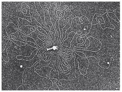
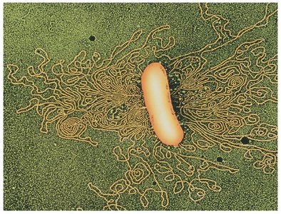
Bacterial DNA Compaction
Bacterial DNA is associated with DNA-binding proteins such as HU and H-NS (Histone-like Nucleoid Structuring Protein).
These proteins fold and bend DNA, creating coils that compact the DNA within the nucleoid.
Mitochondrial and Chloroplast DNA
Characteristics and Evolutionary Origins
Mitochondria and chloroplasts contain their own DNA, which is inherited maternally in most organisms.
The DNA in these organelles is structurally similar to that of viruses and bacteria, supporting the endosymbiotic theory of their evolutionary origins.
Mitochondrial DNA (mtDNA)
Exists as a double-stranded closed circle in most eukaryotes.
Lacks chromosomal proteins and contains few or no introns.
Gene repetition is rare.
Replication of mtDNA depends on enzymes encoded by nuclear DNA.
Chloroplast DNA (cpDNA)
Chloroplasts provide photosynthetic function in plants.
cpDNA is circular, double-stranded, and lacks the associated proteins found in eukaryotic nuclear DNA.
cpDNA is larger than mtDNA, contains more genes, and includes both introns and gene duplications.
Specialized Chromosomes: Polytene and Lampbrush Chromosomes
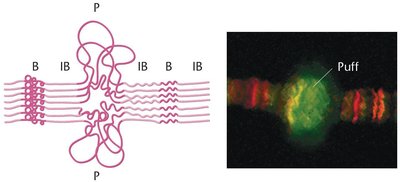
Polytene Chromosomes
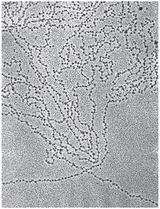
Polytene chromosomes are giant chromosomes found in certain tissues, such as the salivary glands of Drosophila larvae. They are visible in interphase nuclei and represent paired homologs that have undergone multiple rounds of DNA replication without cell division.
Characterized by distinct banding patterns called chromomeres.
Puff regions indicate areas of active transcription, where the DNA is locally uncoiled.
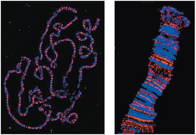
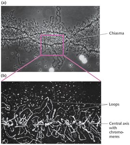
Lampbrush Chromosomes
Lampbrush chromosomes are large meiotic chromosomes with extensive DNA looping, first studied in oocytes of sharks and found in most vertebrate oocytes and some insect spermatocytes. They are easily isolated during the diplotene stage of prophase I of meiosis and contain many condensed chromomeres.
Chromatin Structure in Eukaryotes
Chromatin Organization
During interphase, eukaryotic chromosomes are uncoiled and decondensed into chromatin, which is dispersed throughout the nucleus and replicated.
During cell division, chromatin condenses into visible chromosomes.
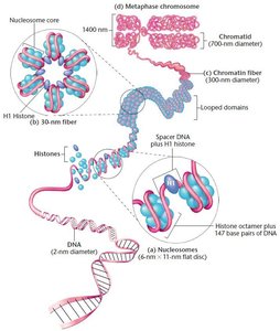
Histones and Nucleosomes
Chromatin is associated with histones, which are positively charged proteins that bind to DNA via electrostatic interactions with the negatively charged phosphate backbone.
There are five main types of histones, all rich in lysine and arginine.
Chromatin fibers are composed of a linear array of spherical particles called nucleosomes.
Each nucleosome core particle consists of histones H2A, H2B, H3, and H4, forming a tetrameric structure around which DNA is wrapped.
Chromatin Remodeling and Histone Modifications
Chromatin Remodeling
Chromatin remodeling is a key process in epigenetics, allowing DNA to become accessible for replication, repair, and gene expression.
Remodeling involves relaxing the compact chromatin structure and providing mechanisms to reverse inactivity.
X-ray diffraction studies have revealed the superhelical structure of DNA wrapped around histones, forming the principal packaging unit in the eukaryotic nucleus.
Histone Tails and Their Role
Unstructured histone tails protrude from the nucleosome core and interact with adjacent nucleosomes, influencing chromatin compaction and accessibility.
These tails are subject to various post-translational modifications (e.g., acetylation, methylation, phosphorylation) that regulate gene expression and DNA accessibility.
