 Back
BackChromosome Structure and Organization in Prokaryotes and Eukaryotes
Study Guide - Smart Notes
 Tailored notes based on your materials, expanded with key definitions, examples, and context.
Chromosome Structure and Organization
Introduction to Chromosome Structure
Chromosomes are highly organized structures composed of DNA and associated proteins. Their structure and organization differ significantly between prokaryotes and eukaryotes, reflecting differences in genome size, complexity, and cellular compartmentalization.
Genome: The total genetic material in an organism.
Genotype: The genetic constitution of an organism.
Chromatin: The complex of DNA and proteins (mainly histones) found in eukaryotic nuclei.
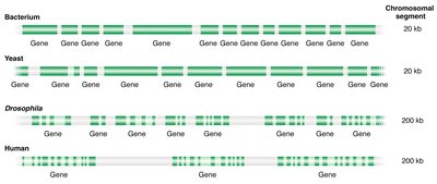
Gene Organization in Prokaryotes and Eukaryotes
Gene Structure and Arrangement
Genes are segments of DNA that encode functional products, typically proteins. The organization of genes varies between prokaryotes and eukaryotes:
Prokaryotic Genes: Generally lack introns, are closely packed, and often organized in operons.
Eukaryotic Genes: Usually contain introns and exons, are separated by large intergenic regions, and are regulated by complex regulatory sequences.
Key regions in all protein-coding genes:
Coding region: Contains the information for the structure of the expressed protein.
Regulatory region: Contains information on where and when a gene will be transcribed; includes the promoter.
Transcription termination sequence: Signals where transcription should end.
Gene Density and Non-coding DNA
Gene density is higher in prokaryotes than in eukaryotes. Eukaryotic genomes contain large amounts of non-coding DNA, including introns and intergenic regions.
Introns: Non-coding sequences within genes, removed during mRNA processing (mainly in eukaryotes).
Intergenic regions: Non-coding DNA between genes.
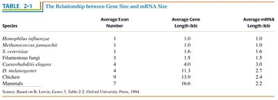
Relationship Between Gene Size and mRNA Size
Gene size and mRNA size are related, but the presence of introns in eukaryotes means that genes are often much larger than the mRNAs they produce.
Species | Average Exon Number | Average Gene Length (kb) | Average mRNA Length (kb) |
|---|---|---|---|
Hemophilus influenzae | 1 | 1.0 | 1.0 |
Methanococcus jannaschii | 1 | 1.0 | 1.0 |
S. cerevisiae | 1 | 1.6 | 1.6 |
Filamentous fungi | 3 | 5.5 | 1.5 |
Caenorhabditis elegans | 4 | 6.5 | 2.0 |
D. melanogaster | 4 | 11.3 | 2.7 |
Chicken | 9 | 13.9 | 2.2 |
Mammals | 7 | 16.6 | 2.2 |
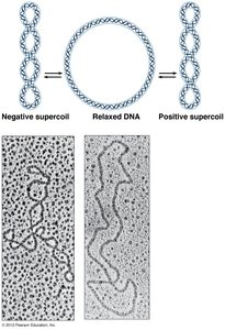
Chromosome Packaging and Compaction
Prokaryotic Chromosome Structure
Prokaryotic chromosomes are typically circular and found in the nucleoid region of the cell. They are compacted by supercoiling and the action of histone-like proteins.
Supercoiling: The coiling of the DNA molecule to reduce its volume and fit within the cell.
Nucleoid: The region in a prokaryotic cell where the chromosome is located.
Eukaryotic Chromosome Structure
Eukaryotic chromosomes are linear and found in the nucleus. DNA is wrapped around histone proteins to form nucleosomes, which further fold into higher-order structures, resulting in a high degree of compaction.
Nucleosome: The basic unit of DNA packaging, consisting of DNA wrapped around a histone octamer.
Chromatin: The complex of DNA and proteins that forms chromosomes.
Metaphase chromosome: The most condensed form of chromatin, visible during cell division.
Chromosome Territories and Nuclear Organization
Chromosome Territories
During interphase, each chromosome occupies a distinct region within the nucleus, known as a chromosome territory. This organization is important for gene regulation and genome stability.
Chromosomes are not randomly distributed but are confined to specific nuclear sub-regions.
Chromosome territories are dynamic and can influence gene expression.
Types of Chromatin
Euchromatin and Heterochromatin
Chromatin exists in two main forms:
Euchromatin: Less condensed, transcriptionally active, and stains lightly with Giemsa.
Heterochromatin: Highly condensed, transcriptionally inactive, and stains darkly with Giemsa.
Heterochromatin can be further divided into:
Constitutive heterochromatin: Always condensed and generally contains repetitive DNA.
Facultative heterochromatin: Can switch between condensed and relaxed states depending on gene expression needs.
Chromosome Banding and Karyotyping
Giemsa Banding (G-banding)
Giemsa staining is used to visualize chromosomes during metaphase, producing characteristic banding patterns that help identify individual chromosomes and detect abnormalities.
AT-rich regions stain more darkly (heterochromatin).
GC-rich regions stain lightly (euchromatin).
Chromosomal Abnormalities and Disease
Philadelphia Chromosome and Chronic Myeloid Leukemia (CML)
Chromosomal rearrangements, such as translocations, can lead to diseases like cancer. The Philadelphia chromosome is a shortened chromosome 22 resulting from a translocation with chromosome 9, commonly found in CML patients.
Identified by Giemsa and fluorescent staining techniques.
Serves as a molecular marker for diagnosis and treatment monitoring.
Summary Table: Key Differences Between Prokaryotic and Eukaryotic Chromosomes
Feature | Prokaryotes | Eukaryotes |
|---|---|---|
Chromosome shape | Circular | Linear |
Location | Nucleoid (cytoplasm) | Nucleus |
Packaging proteins | Histone-like proteins | Histones |
Gene density | High | Low |
Introns | Rare | Common |
Chromatin structure | Not present | Present (chromatin) |
