 Back
Chromosome Structure and Organization in Prokaryotes and Eukaryotes
Study Guide - Smart Notes
 Tailored notes based on your materials, expanded with key definitions, examples, and context.
Chromosome Structure
Abbreviations for DNA/RNA Length
In genetics, the length of DNA or RNA molecules is commonly described using the following abbreviations:
bp (base pair): One base pair, approximately 3.4 Å (340 pm) in length along the strand.
kb (kilobase pair): 1,000 base pairs.
Mb (megabase pair): 1,000,000 base pairs.
Gb (gigabase pair): 1,000,000,000 base pairs.
Viral and Bacterial Chromosomes
General Features
Viral and bacterial chromosomes are structurally simpler than those of eukaryotes:
Composed of a single nucleic acid molecule (either DNA or RNA).
Largely devoid of associated proteins.
Much smaller in size and contain less genetic information than eukaryotic chromosomes.
Bacterial Chromosome Structure
Bacterial chromosomes are typically:
Circular and double-stranded DNA.
Compacted into a region called the nucleoid.
Associated with DNA-binding proteins such as HU and H-NS (Histone-like Nucleoid Structuring Protein), which help fold and bend DNA for compaction.
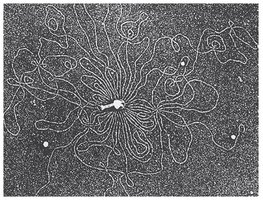
Key Features of Bacterial Chromosomes
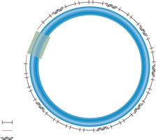
Most are circular and usually a few million base pairs in length.
Typically, a single type of chromosome is present, but it may exist in multiple copies.
Several thousand different genes are interspersed throughout the chromosome.
Regions between adjacent genes are called intergenic regions.
One origin of replication is required to initiate DNA replication.
Repetitive sequences may be interspersed throughout the chromosome.
Bacterial Chromosome Localization
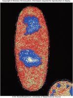
The bacterial chromosome is found in the nucleoid region of the cell, which is not surrounded by a membrane.
Eukaryotic Chromosomes
General Organization
Eukaryotic chromosomes are long, linear DNA molecules that require three types of DNA sequences for proper replication and segregation:
Origins of replication: Multiple origins per chromosome, spaced about every 100,000 base pairs.
Centromeres: Required for proper segregation during mitosis and meiosis; form recognition sites for kinetochore proteins.
Telomeres: Specialized sequences at the ends of linear chromosomes that prevent chromosome shortening and sticky ends.
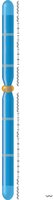
Key Features of Eukaryotic Chromosomes
Range from tens of millions to hundreds of millions of base pairs in length.
Occur in sets (e.g., diploid organisms have two sets).
Genes are interspersed throughout the chromosome.
Contain many origins of replication, centromeres, and telomeres.
Repetitive sequences are commonly found near centromeric and telomeric regions, but may also be interspersed throughout the chromosome.
Comparison: Eukaryotic vs. Prokaryotic Chromosomes
Eukaryotic chromosomes are linear, larger, and have multiple origins of replication, centromeres, and telomeres.
Prokaryotic chromosomes are typically circular, smaller, and have a single origin of replication without centromeres or telomeres.
Genome Size Variation
Genome Sizes in Eukaryotes
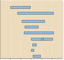
Eukaryotic genome sizes vary widely among different groups of organisms, from fungi to mammals and salamanders.
Examples: Salamander Genome Sizes
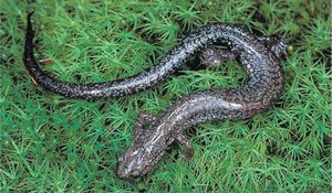
Some species, such as Plethodon larselli and Plethodon richmondi, show dramatic differences in genome size, with P. larselli having more than twice the genome size of P. richmondi.

Composition of the Human Genome
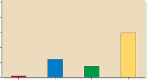
The human genome is composed of:
2% protein-coding regions (exons)
24% introns and other parts of genes
15% unique noncoding DNA
59% repetitive DNA
Chromatin Structure and Compaction
Chromatin and Nucleosomes
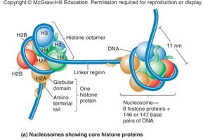
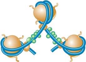
DNA in eukaryotic cells is tightly compacted with proteins to form chromatin. The basic repeating unit of chromatin is the nucleosome, which consists of double-stranded DNA wrapped around an octamer of histone proteins (two copies each of H2A, H2B, H3, and H4).
Each nucleosome contains about 146 base pairs of DNA making 1.65 negative superhelical turns around the histone octamer.
Histones are rich in positively charged amino acids, facilitating their interaction with negatively charged DNA.
Beads-on-a-String Structure
Nucleosomes appear as "beads on a string" under the electron microscope, with linker DNA connecting adjacent nucleosomes. This structure shortens the DNA length about seven-fold.
30-nm Fiber
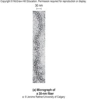
Nucleosomes further associate to form a more compact structure known as the 30-nm fiber, which shortens the DNA about seven-fold beyond the nucleosome level.
Higher-Order Chromatin Structure
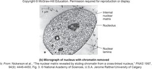
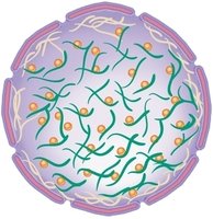
Beyond the 30-nm fiber, chromatin is further compacted by interactions with the nuclear matrix, which consists of the nuclear lamina (lining the inner membrane) and the internal nuclear matrix (filling the nucleus interior). This compaction organizes DNA into radial loop domains.
Matrix-Attachment Regions (MARs)
MARs (or SARs) are DNA sequences that anchor chromatin loops to the nuclear matrix, forming radial loops of 25,000 to 200,000 base pairs.

Euchromatin vs. Heterochromatin
Euchromatin: Less condensed, transcriptionally active, regions where the 30-nm fiber forms radial loop domains.
Heterochromatin: Highly compacted, transcriptionally inactive, radial loop domains compacted even further.

Metaphase Chromosomes
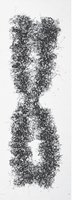
During mitosis, chromatin compaction increases dramatically. By the end of prophase, sister chromatids are entirely heterochromatic, forming the most tightly packed chromatin structure.
Chromatin Remodeling and Chemical Modifications
Chromatin Remodeling
Chromatin structure can be dynamically modified by:
Chromatin remodeling complexes
Chemical modifications on histone tails, including acetylation, methylation, and phosphorylation
DNA methylation (e.g., 5-methyl cytosine)
These modifications regulate gene expression by altering the accessibility of DNA to transcription machinery.
