 Back
BackChromosome Structure, Banding, and Abnormalities in Eukaryotes
Study Guide - Smart Notes
 Tailored notes based on your materials, expanded with key definitions, examples, and context.
Chromosomal Theory of Inheritance
Fundamental Principles
The chromosomal theory of inheritance states that inherited traits are controlled by genes located on chromosomes, which are faithfully transmitted from one generation to the next. This theory forms the basis for understanding genetic inheritance and chromosomal behavior during cell division.
Genes are segments of DNA located on chromosomes.
Chromosomes are the carriers of genetic information.
Transmission of chromosomes ensures inheritance of traits.
Cytogenetics
Definition and Techniques
Cytogenetics is a branch of genetics that studies the relationship between chromosomes and cell behavior, especially during mitosis and meiosis. It utilizes several techniques to analyze chromosome structure and function.
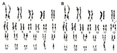
Karyotyping: Visualization of the complete set of chromosomes in a cell.
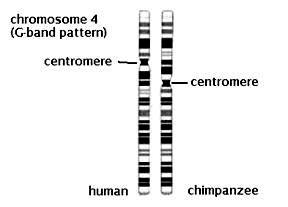
G-banding: Chromosomes are stained with Geimsa stain to reveal unique banding patterns.
Molecular cytogenetics: Includes techniques like Fluorescent in situ hybridization (FISH) and Comparative Genomic Hybridization (CGH).
FISH: Uses fluorescent probes to detect specific DNA sequences.
CGH: Analyzes copy number variations (CNVs) relative to ploidy level.
Chromosome Staining and Banding Patterns
G-Banding Procedure
G-banding is a technique used to distinguish chromosomes based on their banding patterns. Chromosomes are synchronized in culture, halted in metaphase, digested with trypsin, and stained with Geimsa.
Heterochromatic regions: Gene-poor, produce dark G bands.
Euchromatic regions: Gene-rich, produce light G-negative bands.
Bands are numbered from the centromere to the telomere for precise identification.
Chromosome Morphology
Types of Chromosomes
Chromosomes are classified based on the position of the centromere:
Metacentric: Centromere is in the middle.
Sub-metacentric: Centromere is slightly off-center.
Acrocentric: Centromere is near one end.
Telocentric: Centromere is at the end (not found in humans).
Chromosomal Composition and Ploidy
Human Chromosome Sets
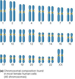
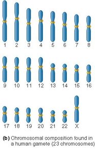
Human somatic cells are diploid (2n), containing 46 chromosomes arranged in 23 homologous pairs. Gametes are haploid (n), containing 23 chromosomes.
Diploid (2n): Two chromosomes per set.
Haploid (n): One chromosome per set.
Gametes: Sperm and egg cells are haploid.
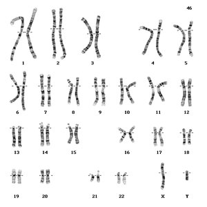
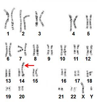
Karyotyping and Chromosome Abnormalities
Diagnostic Applications
Karyotyping is used to detect chromosomal abnormalities, including structural and numerical changes. Chromosome abnormalities can occur during gametogenesis or embryogenesis.
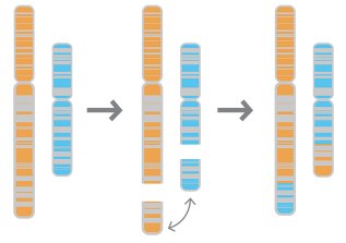
Structural abnormalities: Affect chromosome morphology (e.g., deletions, duplications, translocations, inversions).
Numerical abnormalities: Affect chromosome ploidy (e.g., monosomy, trisomy).
Structural Chromosome Abnormalities
Types and Examples
Structural abnormalities include:
Duplication: Extra genetic material.
Deletion: Loss of genetic material.
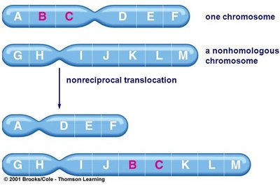
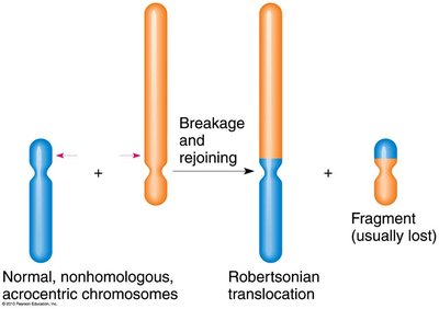
Translocation: Exchange of material between chromosomes (reciprocal or non-reciprocal).
Inversion: Segment of chromosome is reversed.
Chromosome instability syndrome: Leads to chromosomal breakage.
Conditions Caused by Structural Abnormalities
Condition | Frequency | Syndrome | Characteristics |
|---|---|---|---|
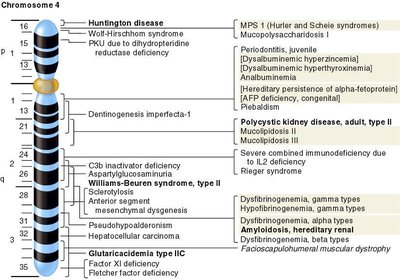
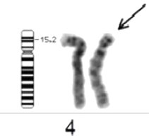
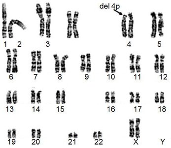
Deletion of p arm of chromosome 4 | 1/50,000 births | Wolf-Hirschhorn | Delayed growth, seizures, intellectual disability, facial features |
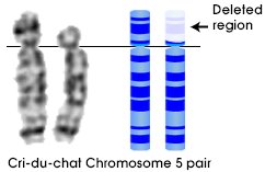
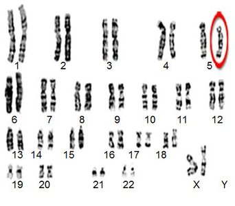
Deletion of p arm of chromosome 5 | 1/50,000 births | Cri-du-chat | Cognitive, speech, motor delays, behavioral problems, facial features |
Duplication of p arm of chromosome 17 | 1/2,500 births | Charcot-Marie-Tooth | Loss of sensation, muscular contractions, skeletal deformation |
Wolf-Hirschhorn Syndrome (Chromosome 4 Deletion)
Prominent forehead, wideset eyes, broad-beaked nose, mental retardation, seizures, heart and muscle defects.
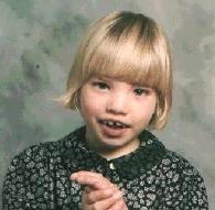
Cri-du-chat Syndrome (Chromosome 5 Deletion)
Microcephaly, round face, small chin, eyes far apart, skin folds, small nose bridge, heart defects, behavioral problems, mental retardation.
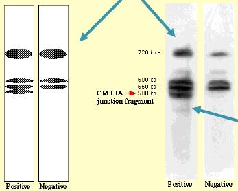
Charcot-Marie-Tooth Disease (Chromosome 17 Duplication)
Duplication of PMP22 gene on chromosome 17.
Large duplications visible under microscope; small duplications detected by gel electrophoresis.
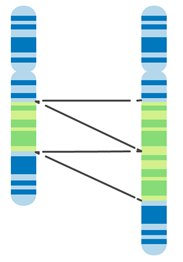
Translocations
Reciprocal (Balanced) Translocation
Equal exchange of chromosomal material between two chromosomes.
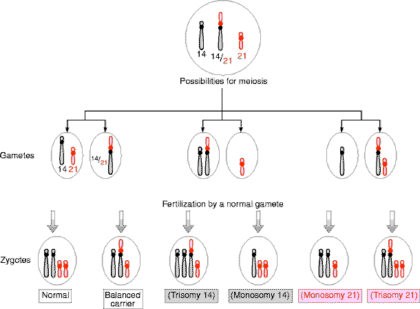
Non-Reciprocal (Robertsonian) Translocation
Occurs when breaks occur at the ends of two non-homologous acrocentric chromosomes, which are then joined together. Familial Down syndrome is an example.

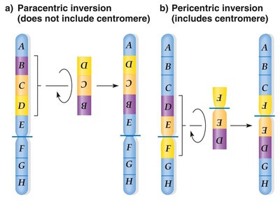
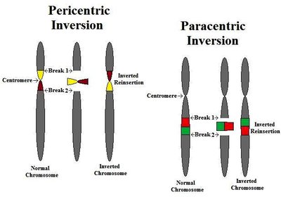
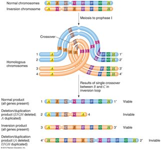
Chromosomal Inversions
Types of Inversions
Inversions are classified as paracentric (does not include centromere) or pericentric (includes centromere).
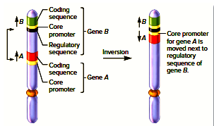
Inversions do not necessarily block transcription.
Can affect gene regulation if regulatory sequences are moved.
Numerical Chromosomal Abnormalities
Ploidy and Somy
Numerical abnormalities affect chromosome sets. If all sets are affected, ploidy changes (e.g., 2n, 3n, 4n). If only one set is affected, the condition is called a somy (e.g., 2n+1).
Aneuploidy: Gain or loss of one or more chromosomes.
Monosomy: Loss of a single chromosome.
Trisomy: Gain of a single chromosome.
Tetrasomy: Gain of two chromosomes.
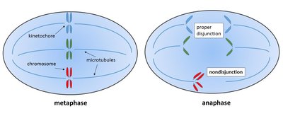
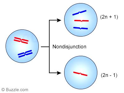
Non-Disjunction
Non-disjunction is the failure of chromosomes to separate properly during cell division, resulting in mosaicism or aneuploidy.
Can occur in mitosis or meiosis.
Mosaicism: Some cells are normal, others are aneuploid.
Pallister-Killian Syndrome
Duplication of p arm of chromosome 12.
Condition is usually mosaic; developmental disability, epilepsy.
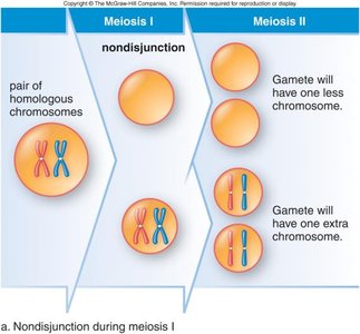
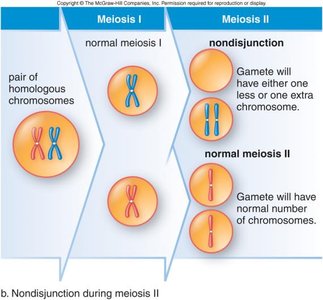
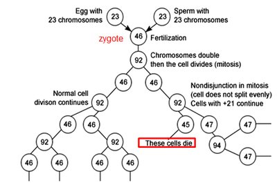
Non-Disjunction During Meiosis
Non-disjunction during meiosis leads to gametes with abnormal chromosome numbers, resulting in aneuploid offspring.
Aneuploid Conditions in Humans
Autosomal Aneuploidy
Condition | Frequency | Syndrome | Characteristics |
|---|---|---|---|
Trisomy 21 | 1/800 | Down | Mental retardation, abnormal palm creases, slanted eyes, flattened face, short stature |
Trisomy 18 | 1/6000 | Edwards | Mental and physical retardation, facial abnormalities, extreme muscle tone, early death |
Trisomy 13 | 1/15000 | Patau | Mental and physical retardation, organ defects, triangular nose, early death |
Sex Chromosome Aneuploidy
Condition | Frequency | Syndrome | Characteristics |
|---|---|---|---|
XXY; XXXY; XXXXY | 1/1000 (males) | Klinefelter | Sexual immaturity, breast swelling |
XYY | 1/1000 (males) | Jacobs | Tall |
XXX | 1/1500 (females) | Triple X | Tall and thin, menstrual irregularity |
XO | 1/5000 (females) | Turner | Short stature, webbed neck, sexually undeveloped |
Prenatal Tests for Chromosomal Defects
Diagnostic Methods
Amniocentesis: Fluid surrounding fetus is sampled for chromosomal analysis.
Chorionic villus sampling (CVS): Biopsy of placental cells.
Cellular mitotic analysis: Used with amniocentesis and CVS.
Ploidy and Polyploidy
Definitions
Ploidy: Number of complete sets of chromosomes in a cell.
Haploid: n chromosomes per cell.
Diploid: 2n chromosomes per cell.
Euploidy: Multiple of the normal haploid set.
Polyploidy: More than two chromosomes per set (e.g., triploid, tetraploid).
Aneuploidy: Abnormal number within one or more sets (e.g., 2n+1, 2n-1).
Origins of Polyploidy
Mechanisms
Chromosomal nondisjunction in meiosis produces gametes with abnormal sets.
Fusion of gametes with abnormal chromosome number.
Polyploidy in Plants
Endoreduplication
Endoreduplication (endoreplication or endocycling) is the replication of the nuclear genome without cytokinesis, leading to polyploidy in plants.
Alloploidy vs Autoploidy
Definitions
Alloploid: Hybrid of two different species with more than two sets of chromosomes.
Autoploid: Fusion of gametes from the same species with unreduced chromosomes (2n + 2n = 4n).
Summary Table: Chromosome Structural Abnormalities
Type | Description | Example |
|---|---|---|
Deletion | Loss of genetic material | Wolf-Hirschhorn, Cri-du-chat |
Duplication | Extra genetic material | Charcot-Marie-Tooth |
Translocation | Exchange between chromosomes | Robertsonian, Reciprocal |
Inversion | Segment reversed | Paracentric, Pericentric |
Additional info: Academic context was added to clarify definitions, mechanisms, and diagnostic methods. Tables were reconstructed for clarity and completeness. Images were included only when directly relevant to the explanation.
