 Back
BackDNA Dynamics and Chromatin Packaging: Structure, Topology, and Organization in Eukaryotes
Study Guide - Smart Notes
 Tailored notes based on your materials, expanded with key definitions, examples, and context.
DNA Topology and Supercoiling
Forms of DNA Topology
DNA molecules can adopt several topological forms, which influence their biological functions and interactions with proteins. The main forms include relaxed, linear, and supercoiled DNA.
Relaxed DNA: The natural state of DNA with no additional twisting beyond the double helix.
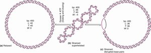
Supercoiled DNA: DNA that is twisted further than its relaxed state. Supercoiling can be negative (underwound, facilitating strand separation) or positive (overwound, resisting unwinding).
Linear DNA: DNA with open ends, typical of eukaryotic chromosomes.
Cellular DNA: Most cellular DNA is negatively supercoiled, which aids in processes such as replication and transcription by making strand separation easier.
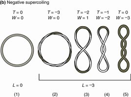
Key Topological Terms
Linking Number (L): The total number of times one DNA strand wraps around the other in a closed DNA molecule.
Twist (T): The number of helical turns in the DNA double helix.
Writhe (W): The number of supercoils or the coiling of the double helix upon itself.
The relationship is given by the equation:
Biological Importance of Supercoiling
Negative supercoiling underwinds DNA, making it easier for enzymes to separate the strands during replication and transcription.
Positive supercoiling overwinds DNA, making strand separation more difficult.
Topoisomerases: Enzymes Managing DNA Topology
General Function
Topoisomerases are enzymes that relieve torsional strain in DNA by cutting, unwinding, and resealing DNA strands. They are essential for preventing DNA from becoming overwound or tangled during replication and transcription.
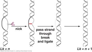
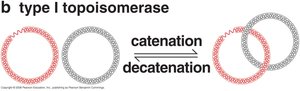
Type I Topoisomerase
Cuts one strand of the DNA double helix (nick).
Allows the uncut strand to rotate through the break, relieving supercoiling tension.
Reseals the strand after relaxation.
Changes the linking number (Lk) by +1 or –1.
Does not require ATP; energy from the broken phosphodiester bond is conserved and reused.
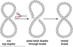
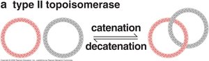
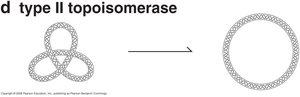
Type II Topoisomerase
Cuts both strands of one DNA double helix.
Allows another segment of the DNA helix to pass through the break, untangling or relaxing the molecule.
Reseals both strands after passage.
Changes the linking number (Lk) by ±2.
Requires ATP to drive the movement of DNA segments.
Topoisomerases and DNA Untangling
Topoisomerases can catenate (link) and decatenate (unlink) circular DNA molecules.
They also relax DNA knots, ensuring proper chromosome segregation and function.
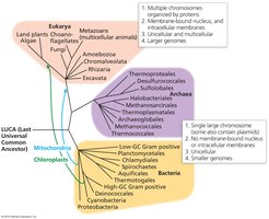
DNA Organization Across Domains of Life
Comparative Overview
Bacteria: Usually possess a single circular chromosome and plasmids, lack a nucleus, and have smaller genomes.
Archaea: Typically have a single circular chromosome, share features with both bacteria and eukaryotes, and have intermediate complexity.
Eukaryotes: Contain multiple linear chromosomes organized by histone proteins, housed within a membrane-bound nucleus, and have larger genomes.
Eukaryotic DNA Organization and Chromatin Structure
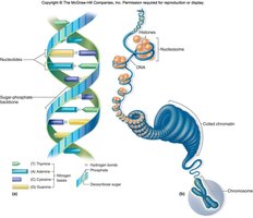
Hierarchical DNA Packaging
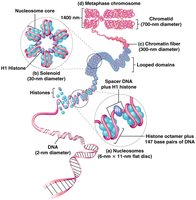
Eukaryotic DNA is highly organized to fit within the nucleus. The packaging involves several hierarchical levels:
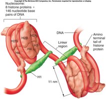
Nucleosome: The basic unit of chromatin, consisting of DNA wrapped around a histone octamer (two each of H2A, H2B, H3, and H4).
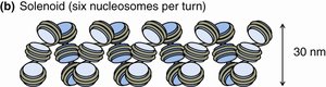
Chromatin Fiber: Nucleosomes are further coiled into a 30 nm fiber (solenoid), which is then organized into loops and higher-order structures.
Chromosome: The most condensed form, visible during cell division.
Nucleoprotein Complex (Chromatin)
Histones: Package and organize DNA into nucleosomes.
Nonhistone proteins: Regulate gene expression, replication, and chromatin remodeling.
Key Terms:
Nucleosome: DNA wrapped around a histone core.
Histone octamer: Core complex of 8 histones (2 each of H2A, H2B, H3, H4).
Linker DNA: Short DNA segment connecting nucleosomes.
Histone H1: Binds to linker DNA, stabilizing and compacting chromatin.
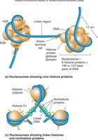
Structure of Eukaryotic Chromosomes
Pairs of histones form an octameric core, around which ~146 base pairs of DNA are wound.
Nucleosomes are connected by linker DNA and stabilized by histone H1.
These filaments fold into loops attached to the nuclear matrix, organizing the genome within the nucleus.
Beads-on-a-String Chromatin
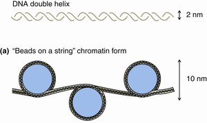
The "beads-on-a-string" model describes nucleosomes connected by linker DNA, representing the least condensed chromatin form (10 nm fiber). This structure is typical of euchromatin, where DNA is accessible for transcription and replication.
Higher-Order Chromatin Organization
The 10 nm fiber coils into a 30 nm chromatin fiber (solenoid), increasing compaction.
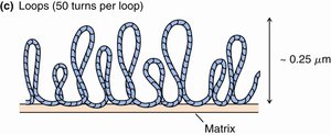
Chromatin fibers form loops anchored to the nuclear matrix, further organizing DNA.
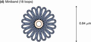
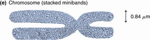
Loops are arranged into minibands, which are stacked to form chromatids and, ultimately, chromosomes.
Fully condensed chromosomes are visible during mitosis, ensuring accurate DNA segregation.
Levels of DNA Organization in Eukaryotic Cells
DNA is packaged in successive levels to fit inside the nucleus, balancing compaction with accessibility for replication and gene expression. The hierarchy is as follows:
Level | Description |
|---|---|
DNA | Double helix (2 nm diameter) |
Nucleosome | "Beads-on-a-string" (10 nm fiber) |
30 nm Fiber (Solenoid) | Coiled nucleosomes |
Looped Domains | Loops anchored to nuclear matrix |
Miniband | 18 loops arranged radially |
Chromatid | Stacked minibands |
Chromosome | Two sister chromatids (most condensed state) |
Summary Table: Key Features of DNA Packaging
Structure | Diameter | Key Components | Function |
|---|---|---|---|
DNA Double Helix | 2 nm | Nucleotides | Genetic information storage |
Nucleosome | 10 nm | Histone octamer + DNA | Basic chromatin unit |
Solenoid Fiber | 30 nm | Coiled nucleosomes | Intermediate compaction |
Looped Domains | ~300 nm | Chromatin loops | Genome organization |
Miniband | ~0.84 µm | 18 loops | Higher-order structure |
Chromatid | 700 nm | Stacked minibands | Chromosome arm |
Chromosome | 1400 nm | Two chromatids | Genetic segregation |
Additional info: Chromatin structure is dynamic and can be remodeled to regulate gene expression, DNA replication, and repair. The accessibility of DNA is modulated by histone modifications and chromatin remodeling complexes.
