 Back
BackDNA Organization in Chromosomes: Structure, Compaction, and Functional Implications
Study Guide - Smart Notes
 Tailored notes based on your materials, expanded with key definitions, examples, and context.
Chromosome Structure in Eukaryotes
Overview of Chromosomes
Chromosomes are highly organized structures composed of DNA and proteins, essential for the storage, expression, and transmission of genetic information. In diploid organisms such as humans, each cell contains two copies of each chromosome (2n = 46), with the total DNA content exceeding 2 meters in length if stretched out. The cell compacts this DNA efficiently to fit within the nucleus.
Diploid genome: Contains two sets of chromosomes, one from each parent.
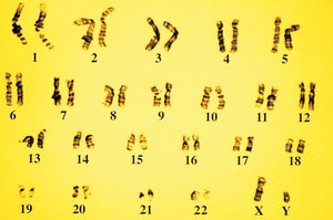
Human karyotype: The complete set of chromosomes in a human cell, visualized for genetic analysis.
Homologous chromosomes: Chromosome pairs with the same genes but possibly different alleles.
Chromatin Composition
Chromatin is the macromolecular complex of DNA and proteins found in the nucleus. It consists of:
DNA: Linear double-stranded molecule encoding genetic information.
Histone proteins: Core proteins (H2A, H2B, H3, H4) around which DNA is wound to form nucleosomes; H1 locks DNA in place.
Non-histone proteins: Include DNA polymerase, scaffold proteins, kinetochore proteins, and transcription factors.
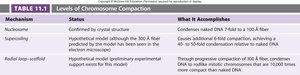
Levels of DNA Compaction
DNA compaction is essential for fitting the genome into the nucleus and for regulating gene expression. Compaction occurs in several hierarchical levels:
Mechanism | Status | What It Accomplishes |
|---|---|---|
Nucleosome | Confirmed by crystal structure | Condenses naked DNA 7-fold to 100 Å fiber |
Supercoiling | Hypothetical model; 300 Å fiber proposed | Causes additional condensation, achieving 40-50 fold compaction |
Radial loop-scaffold | Hypothetical; preliminary experimental support | Progressive compaction into mitotic chromosomes, up to 10,000 times more compact than naked DNA |
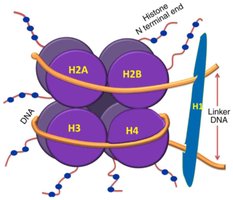
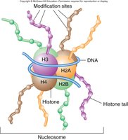
Nucleosome Structure
The nucleosome is the fundamental unit of chromatin structure. It consists of 160 base pairs of DNA wrapped around a histone octamer (two each of H2A, H2B, H3, and H4), with H1 stabilizing the structure. This arrangement resembles a "spool of thread." Nucleosomes are often described as "beads on a string." Their placement along DNA influences gene accessibility and expression.
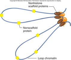
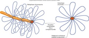
Higher-Order Chromatin Structure
Beyond nucleosomes, chromatin is further compacted by supercoiling and the formation of structural loops. Non-histone proteins (NHPs) bind chromatin and organize it into loops, which are then gathered into rosettes, forming the radial loop-scaffold model. This compaction is essential for chromosome condensation during mitosis.
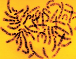
Chromosome Banding Patterns
DNA compaction leads to visible banding patterns on chromosomes, which are used in karyotyping and gene mapping. Dark bands (heterochromatin) are highly compacted and gene-poor, while light bands (euchromatin) are less compact and gene-rich.
Chromatin States: Heterochromatin vs. Euchromatin
Definitions and Properties
Heterochromatin: Highly compacted chromatin, generally transcriptionally inactive, often found at centromeres and telomeres.
Euchromatin: Less compacted chromatin, accessible to transcription machinery, and associated with active gene expression.
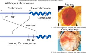
Position Effect Variegation (PEV)
Heterochromatin can spread into adjacent euchromatin, silencing genes in a mosaic pattern. This phenomenon, called position effect variegation, demonstrates the dynamic nature of chromatin states and their impact on gene expression.
Histone Modifications and Chromatin Remodeling
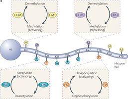
Histone Tail Modifications
Histone proteins have N-terminal tails that extend from the nucleosome and can be covalently modified. These modifications are reversible and include acetylation, methylation, phosphorylation, and ubiquitination. They regulate chromatin structure and gene expression by altering nucleosome interactions and recruiting chromatin modifier proteins.
Types of Histone Modifications
Acetylation: Addition of acetyl groups by histone acetyltransferases (HATs) neutralizes positive charges, loosening chromatin and promoting gene expression. Histone deacetylases (HDACs) reverse this process.
Methylation: Addition of methyl groups by histone methyltransferases (HMTs) can either activate or repress gene expression, depending on the specific amino acid residue modified. Demethylases (HDMs) remove methyl groups.
Phosphorylation: Addition of phosphate groups, often associated with chromatin condensation during mitosis.
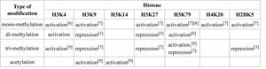
Histone Code
The combination of histone modifications constitutes a "histone code" that determines chromatin state and gene activity. Specific patterns of modifications are associated with either euchromatin (active) or heterochromatin (repressed) states.
Type of modification | H3K4 | H3K9 | H3K14 | H3K27 | H3K79 | H4K20 | H3R5 |
|---|---|---|---|---|---|---|---|
mono-methylation | activation | activation | activation | repression | activation | activation | repression |
di-methylation | activation | repression | activation | repression | activation | repression | repression |
tri-methylation | activation | repression | activation | repression | activation | repression | repression |
acetylation | activation | activation | activation | activation | activation | activation | activation |
DNA Replication and Chromatin Assembly
Origins of Replication
Eukaryotic chromosomes contain multiple origins of replication (oris) to ensure timely duplication of the genome. Chromatin structure around oris is more open, allowing replication machinery to access DNA.
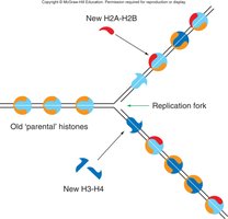
Nucleosome Disassembly and Reassembly
During DNA replication, nucleosomes are temporarily disassembled ahead of the replication fork and reassembled on newly synthesized DNA. New nucleosomes are composed of both recycled and newly synthesized histones, and histone modifications are re-established to maintain epigenetic information.
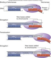
Telomeres and Telomerase
Telomeres are repetitive DNA sequences at the ends of eukaryotic chromosomes that protect against degradation and prevent chromosome fusion. The enzyme telomerase extends telomeric repeats, using an RNA template to add new DNA. Telomerase is active in germ cells, stem cells, and many cancer cells.
Chromosome Segregation: Centromeres and Kinetochores
Centromere Structure and Function
Centromeres are specialized chromosomal regions essential for proper segregation during cell division. They hold sister chromatids together and serve as attachment sites for kinetochore microtubules. In yeast, centromeres contain conserved DNA sequences; in higher eukaryotes, they consist of tandem repeats of non-coding satellite DNA.
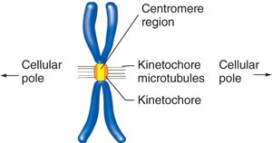
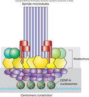
Kinetochore Assembly
Kinetochores are protein complexes that assemble at centromeres, mediating chromosome attachment to spindle microtubules. The histone variant CENP-A replaces H3 in centromeric nucleosomes, providing a scaffold for kinetochore proteins.
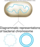
Chromosome Structure in Prokaryotes
Bacterial Chromosomes
Bacteria typically possess a single circular chromosome, ranging from 4 to 5 Mb in size. Unlike eukaryotes, bacterial DNA is not associated with histones but is compacted by supercoiling and nucleoid-associated proteins. The genome is densely packed with genes, and replication initiates from a single origin.
Genome Organization and Diversity
Bacterial genomes are highly efficient, with minimal non-coding DNA. Comparative genomics reveals a core genome shared by all strains and a pangenome encompassing all genes found in different strains. This diversity underlies bacterial adaptability and ecological significance.
Summary Table: Eukaryotic vs. Prokaryotic Chromosome Organization
Feature | Eukaryotes | Prokaryotes |
|---|---|---|
Chromosome number | Multiple, linear | Single, circular |
DNA packaging | Histones, nucleosomes, higher-order structures | Supercoiling, nucleoid-associated proteins |
Origins of replication | Multiple | Single |
Genome size | Large (Mb to Gb) | Small (few Mb) |
Gene density | Lower, with introns and non-coding regions | High, mostly coding DNA |
Additional info: This guide integrates foundational concepts from the study of chromosome structure, chromatin organization, and the molecular mechanisms underlying DNA compaction and segregation, as well as key differences between eukaryotic and prokaryotic chromosome organization.
