 Back
BackDNA Organization in Chromosomes – Study Notes
Study Guide - Smart Notes
 Tailored notes based on your materials, expanded with key definitions, examples, and context.
DNA Organization in Chromosomes
Introduction
The organization of DNA within chromosomes is fundamental to understanding genetic function and regulation. This chapter explores the structural diversity of chromosomes in viruses, bacteria, and eukaryotes, the mechanisms of DNA compaction, and the complexity of eukaryotic genomes.
Viral and Bacterial Chromosomes
Structure and Simplicity
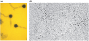
Viral and bacterial chromosomes are typically composed of a single nucleic acid molecule, either DNA or RNA (in viruses), and are largely devoid of the protein scaffolding found in eukaryotic chromosomes.
These chromosomes are much smaller and contain less genetic information than their eukaryotic counterparts.
Viral genetic material can be single- or double-stranded, and either circular or linear.
Viral DNA is inert until it enters a host cell, where it becomes active.
Bacterial Chromosome Structure
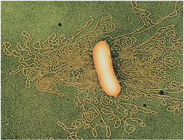
Bacterial chromosomes are typically circular, double-stranded DNA molecules compacted into a region called the nucleoid.
DNA in bacteria is associated with nucleoid-associated proteins (e.g., HU and H-NS) that aid in compaction and organization.
Supercoiling Facilitates DNA Compaction
Supercoiling
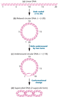
Supercoiling is a process that further compacts DNA, making it more efficient for storage within the cell.
Supercoiled DNA refers to closed-circular DNA molecules that are more compact and sediment more rapidly than linear forms.
Supercoiling is essential for fitting large DNA molecules into small cellular compartments.
Topoisomerases
Topoisomerases are enzymes that cut one or both DNA strands, allowing the helix to be wound or unwound before resealing the ends.
These enzymes are crucial for managing DNA supercoiling during replication and transcription in both prokaryotes and eukaryotes.
Replication and transcription generate supercoils downstream as the double helix unwinds.
Specialized Chromosomes Reveal Variations in DNA Organization
Polytene Chromosomes
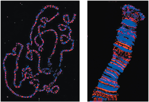
Polytene chromosomes are a unique type of chromosome found in certain tissues of some organisms, such as the salivary glands of Drosophila larvae.
They represent paired homologs that have undergone multiple rounds of DNA replication without cell division (endomitosis).
Polytene chromosomes are visible under light microscopy and display distinct banding patterns.
Puff regions are areas where DNA has uncoiled, indicating high levels of gene activity (transcription).
DNA Organization in Eukaryotes: Chromatin Structure
Chromatin and Histones
During interphase, eukaryotic chromosomes exist as chromatin, a less condensed form that allows access for replication and transcription.
Chromatin consists of DNA wrapped around positively charged proteins called histones (H1, H2A, H2B, H3, H4), which facilitate compaction and regulation.
Electrostatic interactions between histones and the negatively charged DNA backbone are essential for chromatin structure.
Nucleosomes
Nucleosomes are the fundamental repeating units of chromatin, appearing as "beads on a string" under electron microscopy.
Each nucleosome consists of DNA wrapped around a histone octamer, and these structures are further compacted to form chromatids.
Chromatin Remodeling
Chromatin structure must be dynamic to allow DNA-protein interactions necessary for replication and gene expression.
Remodeling involves relaxing the compact structure, exposing DNA regions to regulatory proteins, and reversing these changes during inactivity.
Chemical Modifications of Chromatin
Histone tails are subject to chemical modifications that influence chromatin structure and gene activity:
Acetylation (by histone acetyltransferase, HAT): Addition of acetyl groups to lysine residues neutralizes positive charges, loosening DNA-histone interactions and promoting gene expression.
Methylation (by methyltransferase): Addition of methyl groups to arginine or lysine residues, often associated with gene activation.
Phosphorylation (by kinase): Addition of phosphate groups to serine or histidine residues, affecting chromatin structure and function.
DNA methylation (at CpG sites): Addition of methyl groups to cytosine bases, usually associated with gene silencing.
Euchromatin and Heterochromatin
Euchromatin: Uncoiled, genetically active regions that appear unstained during interphase.
Heterochromatin: Condensed, genetically inactive regions that stain during interphase; includes telomeres (chromosome integrity) and centromeres (chromosome movement).
Chromosome Banding Differentiates Regions along the Mitotic Chromosome
Chromosome Banding Techniques
Banding techniques use differential staining to reveal patterns along the chromosome's length, aiding in chromosome identification and analysis.
C-banding: Stains centromeric heterochromatin.
G-banding: Produces unique banding patterns for each chromosome, allowing for precise identification and comparison.
Eukaryotic Genomes: Repetitive DNA and Sequence Organization
Repetitive DNA Sequences
Eukaryotic genomes contain large amounts of repetitive DNA, which can be categorized as:
Satellite DNA: Highly repetitive, short sequences found in centromeric regions; not present in prokaryotes.
Moderately repetitive DNA: Includes variable number tandem repeats (VNTRs), minisatellites, and microsatellites (short tandem repeats, STRs).
SINEs (Short Interspersed Elements) and LINEs (Long Interspersed Elements): Mobile transposable elements dispersed throughout the genome, constituting over 5% of the human genome.
Pseudogenes and Noncoding DNA
Only 2–10% of the eukaryotic genome encodes proteins.
Pseudogenes are nonfunctional DNA sequences that resemble genes but have accumulated mutations and are not transcribed.
The vast majority of the genome consists of noncoding regions, including regulatory elements, repetitive sequences, and evolutionary remnants.
