 Back
BackDNA Replication and Recombination: Mechanisms and Regulation
Study Guide - Smart Notes
 Tailored notes based on your materials, expanded with key definitions, examples, and context.
DNA Replication and Recombination
Introduction
This chapter explores the molecular mechanisms of DNA replication and recombination, focusing on both prokaryotic and eukaryotic systems. It covers the enzymatic machinery, regulatory processes, and the biological significance of these essential genetic processes.
DNA Is Reproduced by Semiconservative Replication
DNA as a Template
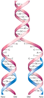
DNA replication relies on the complementarity of nitrogenous bases, allowing each strand to serve as a template for the synthesis of a new complementary strand. This ensures the accurate transmission of genetic information during cell division.
Template strand: The original DNA strand that guides the synthesis of a new strand.
Base pairing: Adenine pairs with thymine, and guanine pairs with cytosine, ensuring fidelity.
Three Modes of DNA Replication
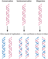
There are three theoretical models for DNA replication:
Semiconservative: Each new DNA molecule consists of one old (parental) and one new strand.
Conservative: The parental double helix remains intact, and an entirely new double helix is synthesized.
Dispersive: Parental DNA is dispersed throughout both strands of the daughter molecules.
Experimental Evidence: Meselson–Stahl Experiment
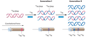
The Meselson–Stahl experiment (1958) provided strong evidence for semiconservative replication in E. coli by using isotopic labeling and density gradient centrifugation.
Method: Cells were grown in heavy nitrogen (15N) and then transferred to light nitrogen (14N). DNA was extracted after each generation and analyzed.
Result: Hybrid DNA molecules after one generation, and a mix of hybrid and light DNA after two generations, supporting the semiconservative model.
Semiconservative DNA Replication in Eukaryotes
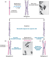
The Taylor–Woods–Hughes experiment (1957) demonstrated semiconservative replication in eukaryotes using Vicia faba (broad bean) and autoradiography to track labeled DNA during cell division.
Autoradiography: A technique that pinpoints the location of radioisotopes in cells, revealing newly synthesized DNA.
Origins, Forks, and Units of Replication
DNA replication begins at specific sites called origins of replication (ORI). The unwinding of the helix at these sites creates replication forks, where DNA synthesis occurs bidirectionally.
Replicon: The length of DNA replicated from a single origin.
Bacterial Replication
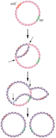
Bacteria typically have a single circular DNA molecule with one origin of replication (OriC). Replication proceeds bidirectionally around the chromosome, forming two replication forks.
DNA Synthesis in Bacteria Involves Five Polymerases and Other Enzymes
DNA Polymerases in Bacteria
Bacterial DNA synthesis involves five DNA polymerases (Pol I, II, III, IV, V), each with specialized functions. DNA polymerase I was the first to be isolated and is essential for DNA repair and primer removal.
Requirements: DNA template, four deoxyribonucleoside triphosphates (dNTPs), and a primer with a free 3'-OH group.
Chain Elongation
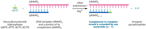
DNA polymerases add nucleotides to the 3' end of a growing DNA strand, releasing pyrophosphate. The reaction proceeds in the 5' to 3' direction.
Equation:
Properties of DNA Polymerases I, II, and III
All three major DNA polymerases can elongate an existing DNA strand but cannot initiate synthesis de novo. They possess exonuclease activity for proofreading and error correction.
Pol I: Removes RNA primers and fills in gaps.
Pol III: Main enzyme for DNA chain elongation.
DNA Polymerase III Holoenzyme
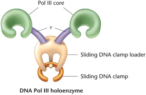
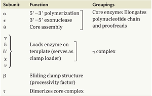
The active form of DNA polymerase III is a holoenzyme, composed of multiple subunits with distinct functions, including polymerization, proofreading, and clamp loading.
Subunit | Function | Groupings |
|---|---|---|
α | 5'→3' polymerization | Core enzyme |
ε | 3'→5' exonuclease | Core enzyme |
θ | Core assembly | Core enzyme |
γ, δ, δ', χ, ψ | Loads enzyme on template (clamp loader) | γ complex |
β | Sliding clamp structure (processivity factor) | |
τ | Dimerizes core complex |
DNA Repair
DNA polymerases also play critical roles in repairing DNA damaged by external factors such as UV light, ensuring genomic stability.
Complex Issues in DNA Replication
Key Issues to Resolve
Seven major challenges must be addressed during DNA replication:
Unwinding of the helix
Relieving supercoiling tension
Synthesizing RNA primers for initiation
Discontinuous synthesis of the lagging strand
Removal of RNA primers
Joining of DNA fragments
Proofreading and error correction
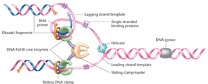
Initiation and Unwinding
DnaA: Initiator protein that binds to OriC, causing DNA to unwind.
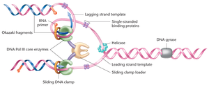
DNA helicase: Unwinds the double helix using energy from ATP hydrolysis.
Single-stranded binding proteins (SSBPs): Stabilize unwound DNA.
DNA gyrase (topoisomerase): Relieves supercoiling ahead of the replication fork.
Primer Synthesis and Elongation
Primase: Synthesizes short RNA primers to provide free 3'-OH ends for DNA polymerase.
RNA priming: Universal mechanism in all domains of life.
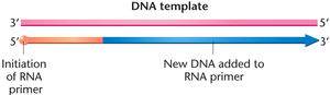
Continuous and Discontinuous Synthesis
Because DNA polymerase can only synthesize in the 5' to 3' direction, replication is continuous on the leading strand and discontinuous on the lagging strand, forming Okazaki fragments.
Leading strand: Synthesized continuously toward the replication fork.
Lagging strand: Synthesized discontinuously away from the fork.
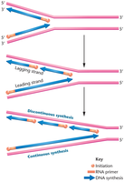
Okazaki Fragments and Ligation
Okazaki fragments: Short DNA segments synthesized on the lagging strand, each initiated by an RNA primer.
DNA ligase: Seals nicks between fragments by forming phosphodiester bonds.
Concurrent Synthesis
Both DNA strands are synthesized simultaneously at the replication fork. The lagging strand forms a loop to allow the DNA polymerase complex to move in the same physical direction as the leading strand.
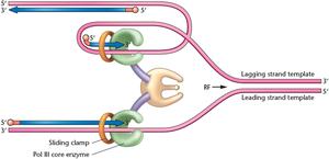
Proofreading
DNA polymerases possess 3' to 5' exonuclease activity, allowing them to remove incorrectly paired nucleotides and enhance replication fidelity.
A Coherent Model of DNA Replication
Summary of Enzymes and Proteins
DNA replication requires the coordinated action of multiple enzymes and proteins, including DNA polymerases, SSBPs, DNA gyrase, DNA helicase, and RNA primase.
Genetic Control of Replication
Gene Mutations Affecting Replication
Mutations in genes encoding replication proteins can disrupt the process, leading to lethal or conditional phenotypes. Examples include ligase-deficient and proofreading-deficient mutations. Temperature-sensitive mutations are expressed only under restrictive conditions.
Gene | Product/Role |
|---|---|
dnaA | Initiator protein |
dnaB | Helicase |
dnaC | Helicase loader |
dnaG | Primase |
lig | DNA ligase |
polA | DNA polymerase I |
polB | DNA polymerase II |
dnaE | DNA polymerase III |
gyrA/B | DNA gyrase |
ssb | Single-stranded binding protein |
uvrD | Helicase II |
recA | Recombination/repair |
Additional info: Table inferred from standard E. coli replication gene functions.
Eukaryotic DNA Replication: Similarities and Differences
General Features
Eukaryotic DNA replication shares core mechanisms with prokaryotes but is more complex due to larger genomes, linear chromosomes, and chromatin structure.
Multiple origins of replication per chromosome
Bidirectional synthesis
Requirement for nucleosome disassembly and reassembly
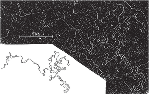
Multiple Origins of Replication
Eukaryotic chromosomes contain many origins of replication, enabling rapid duplication of large genomes. In yeast, these are called autonomously replicating sequences (ARSs).
Prereplication Complex and Initiation
The prereplication complex (pre-RC) assembles at origins during early G1 phase. The origin recognition complex (ORC) marks the site for initiation, ensuring precise timing of replication.
Eukaryotic DNA Polymerases
Multiple DNA polymerases participate in eukaryotic DNA replication:
Pol α: Initiates synthesis and primes both leading and lagging strands.
Pol δ: Synthesizes the lagging strand.
Pol ε: Synthesizes the leading strand.
Other polymerases are involved in DNA repair.
Polymerase switching occurs after primer synthesis, with Pol δ and Pol ε taking over elongation from Pol α.
Replication through Chromatin
DNA is packaged into nucleosomes, which must be temporarily displaced for replication to proceed. Nucleosomes are reassembled on newly synthesized DNA.
Telomeres and the End-Replication Problem
Structure and Function of Telomeres
Telomeres are repetitive DNA sequences at the ends of eukaryotic chromosomes that protect against degradation and fusion. The shelterin complex stabilizes telomere structure.
Telomerase and Telomere Maintenance
Telomerase is a ribonucleoprotein enzyme that extends telomeres by adding repetitive sequences using its RNA component as a template. This compensates for the inability of DNA polymerase to fully replicate chromosome ends.
Active in germ cells, stem cells, and most cancer cells
Inactive in most somatic cells, leading to gradual telomere shortening
Telomerase activity is linked to cellular aging and cancer.
Genetic Recombination
Homologous Recombination
Genetic recombination involves the exchange of DNA between homologous chromosomes, promoting genetic diversity and DNA repair. The process is mediated by specific enzymes and involves several steps:
Endonuclease nicking
Strand displacement and pairing
Ligation
Branch migration
Duplex separation, forming a Holliday junction
Additional info: The Holliday model is a classic representation of homologous recombination.
