 Back
BackDNA Replication in Prokaryotes: Mechanisms and Enzymology (E. coli Model)
Study Guide - Smart Notes
 Tailored notes based on your materials, expanded with key definitions, examples, and context.
DNA Replication: Fundamental Concepts
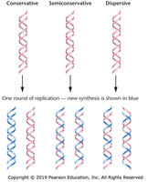
Semiconservative Model of DNA Replication
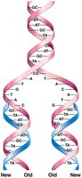
DNA replication is the process by which a cell copies its entire genome before cell division. The semiconservative model, experimentally confirmed by Meselson and Stahl, states that each daughter DNA molecule consists of one parental (old) strand and one newly synthesized strand.
Conservative model: Parental DNA remains intact; new molecule is entirely new DNA.
Semiconservative model: Each daughter DNA has one old and one new strand.
Dispersive model: Parental and new DNA are interspersed in both strands.
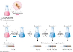
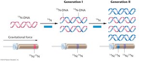
Experimental confirmation: Meselson and Stahl used 15N and 14N isotopes to distinguish old and new DNA, showing intermediate densities after one replication cycle, consistent with semiconservative replication.
Overview of DNA Replication in Prokaryotes
General Features
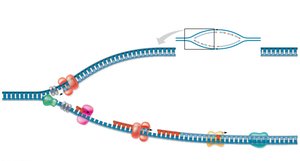
DNA replication is a highly coordinated process involving multiple enzymes and proteins. In E. coli, replication starts at a single origin (oriC) and proceeds bidirectionally, forming two replication forks that move in opposite directions around the circular chromosome.
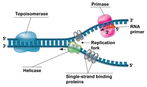
Replication bubble: The region where the double helix is unwound and replication occurs.
Replication fork: The Y-shaped structure where new DNA synthesis takes place.
Bidirectional replication: Two forks move away from the origin, synthesizing DNA in both directions.
Semidiscontinuous synthesis: Leading strand is synthesized continuously; lagging strand is synthesized in short Okazaki fragments.
Initiation of DNA Replication
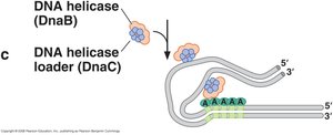
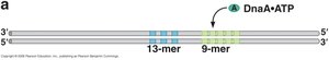
Origin of Replication (oriC) and Initiator Proteins
Replication begins at the oriC locus, which is approximately 245 base pairs long and rich in A–T base pairs, making it easier to unwind. The oriC contains specific sequence repeats:
9-mer repeats: Binding sites for the initiator protein DnaA.
13-mer repeats: A–T-rich regions that are the first to unwind, forming the replication bubble.

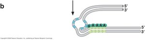
DnaA: Binds to 9-mer repeats, causing local unwinding at 13-mer repeats.
DnaC: Helicase loader, helps load DnaB onto single-stranded DNA.
DnaB: DNA helicase, unwinds the double helix using ATP.
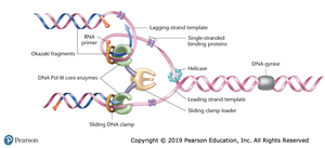
Enzymes and Proteins of the Replication Fork
Key Enzymes and Their Functions
Multiple enzymes and proteins coordinate to ensure accurate and efficient DNA replication:
Helicase (DnaB): Unwinds the DNA double helix ahead of the replication fork.
Single-strand binding proteins (SSBs): Stabilize single-stranded DNA and prevent reannealing.
Topoisomerase (DNA gyrase): Relieves supercoiling ahead of the fork by cutting and resealing DNA strands.
Primase: Synthesizes short RNA primers to provide a 3′-OH group for DNA polymerase.
DNA polymerase III holoenzyme: Main enzyme complex for DNA synthesis; contains two core polymerases, sliding clamps, clamp loader (γ complex), and τ proteins for coordination.
DNA polymerase I: Removes RNA primers and fills gaps with DNA.
DNA ligase: Seals nicks between Okazaki fragments, forming a continuous strand.
Mechanism of DNA Synthesis
Leading and Lagging Strand Synthesis
DNA polymerases can only add nucleotides to the 3′-OH end, so all new DNA is synthesized in the 5′ → 3′ direction. Because the two template strands are antiparallel, synthesis occurs differently on each:
Leading strand: Synthesized continuously toward the replication fork.
Lagging strand: Synthesized discontinuously away from the fork as Okazaki fragments, each starting with an RNA primer.
Trombone model: The lagging strand loops so both polymerases move together in the same overall direction.
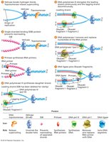
Okazaki Fragment Processing
On the lagging strand, primase periodically synthesizes new RNA primers. DNA polymerase III extends these primers, forming Okazaki fragments. When a fragment is complete, the polymerase releases, and a new clamp is loaded for the next fragment.
RNase H: Removes most of the RNA primer from RNA/DNA hybrids.
DNA polymerase I: Removes remaining ribonucleotides and fills the gap with DNA.
DNA ligase: Seals the final nicks, joining fragments into a continuous strand.
Coordination and Regulation of Replication
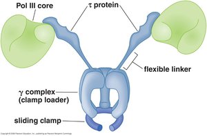
DNA Polymerase III Holoenzyme Structure and Function
The DNA polymerase III holoenzyme is a multi-protein complex that coordinates the synthesis of both DNA strands at the replication fork. It contains:
Two Pol III core enzymes: Each synthesizes one new DNA strand.
Sliding clamps: Encircle DNA, holding polymerase in place for high processivity.
Clamp loader (γ complex): Loads sliding clamps onto DNA at primer-template junctions.
τ proteins: Flexible linkers that coordinate the activities of both polymerases.
Summary Table: Key Enzymes and Their Roles in E. coli DNA Replication
Enzyme/Protein | Function |
|---|---|
DnaA | Binds oriC, initiates strand separation |
DnaB (Helicase) | Unwinds DNA double helix |
DnaC | Loads helicase onto DNA |
SSB | Stabilizes single-stranded DNA |
Topoisomerase (DNA gyrase) | Relieves supercoiling ahead of fork |
Primase | Synthesizes RNA primers |
DNA polymerase III | Main DNA synthesis enzyme |
Sliding clamp | Holds polymerase on DNA |
Clamp loader (γ complex) | Loads sliding clamp onto DNA |
RNase H | Removes RNA primers |
DNA polymerase I | Removes RNA primers, fills gaps with DNA |
DNA ligase | Seals nicks between fragments |
Summary of the Replication Process in E. coli
Initiation: DnaA binds oriC, DnaC loads DnaB (helicase), SSB stabilizes ssDNA, topoisomerase relieves supercoiling.
Elongation: Primase synthesizes RNA primers, clamp loader loads sliding clamps, DNA polymerase III synthesizes leading and lagging strands simultaneously.
Okazaki fragment maturation: RNase H and DNA polymerase I remove RNA primers and fill gaps, DNA ligase seals nicks.
Result: Two identical, semiconservative DNA molecules.
Key Concepts and Applications
Semiconservative replication ensures genetic continuity.
Multiple enzymes and proteins act in a coordinated manner for high fidelity and efficiency.
Understanding prokaryotic replication provides a foundation for studying eukaryotic DNA replication and biotechnological applications.
