 Back
BackDNA Replication: Mechanisms, Enzymes, and Regulation
Study Guide - Smart Notes
 Tailored notes based on your materials, expanded with key definitions, examples, and context.
DNA Replication: Mechanisms, Enzymes, and Regulation
Overview of DNA Replication
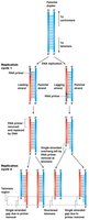
DNA replication is a fundamental process that ensures the accurate duplication of the genetic material in all living organisms. The process is semiconservative and bidirectional, involving a complex interplay of enzymes and regulatory sequences to produce two identical DNA duplexes from one parental molecule.
Semiconservative and Bidirectional Replication
Semiconservative replication: Each daughter DNA molecule consists of one parental (original) strand and one newly synthesized strand.
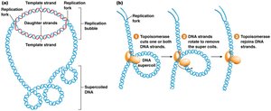
Bidirectional replication: Replication proceeds outward in both directions from the origin, forming replication forks and a replication bubble.
Universality: The general mechanism is conserved across Bacteria, Archaea, and Eukarya, though specific proteins and enzymes may differ.
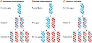
Models of DNA Replication
After the discovery of DNA structure, three models were proposed to explain how DNA is replicated:
Semiconservative model: Each daughter duplex contains one parental and one daughter strand.
Conservative model: One duplex contains both parental strands, the other both daughter strands.
Dispersive model: Each daughter duplex contains interspersed parental and daughter segments.
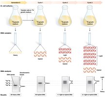
The Meselson-Stahl Experiment
The Meselson-Stahl experiment (1958) provided definitive evidence for the semiconservative model. E. coli were grown in heavy nitrogen (15N), then transferred to light nitrogen (14N). DNA was extracted after each replication cycle and separated by density gradient centrifugation.
After one replication cycle, DNA had intermediate density (14N/15N).
After two cycles, half the DNA was light (14N/14N), half intermediate (14N/15N).
These results supported only the semiconservative model.
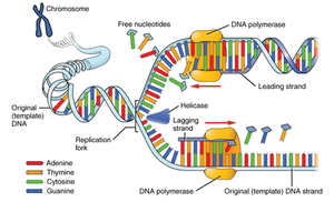
Mechanics of DNA Replication
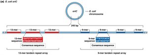
Origin of replication (oriC in E. coli): Specific DNA sequence where replication begins.
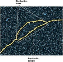
Replication fork: Y-shaped region where the DNA is split into two single strands and copied.
Replication bubble: The region of locally unwound DNA where replication occurs.
Enzymes and Proteins Involved in Replication
Helicase: Unwinds the DNA double helix.
Single-stranded binding proteins (SSB): Stabilize unwound DNA.
Topoisomerase: Relieves supercoiling ahead of the replication fork.
Primase: Synthesizes short RNA primers needed for DNA polymerase to begin synthesis.
DNA polymerase III (bacteria): Main enzyme for DNA synthesis; synthesizes leading and lagging strands.
DNA polymerase I (bacteria): Removes RNA primers and replaces them with DNA.
DNA ligase: Seals nicks between Okazaki fragments on the lagging strand.
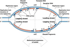
Leading and Lagging Strand Synthesis
DNA polymerase can only synthesize DNA in the 5' to 3' direction, resulting in continuous synthesis on the leading strand and discontinuous synthesis on the lagging strand (Okazaki fragments).
Leading strand: Synthesized continuously toward the replication fork.
Lagging strand: Synthesized discontinuously away from the fork in short Okazaki fragments.
Okazaki fragments: Short DNA segments synthesized on the lagging strand, later joined by DNA ligase.
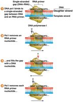
Removal of RNA Primers and Ligation
After DNA synthesis, RNA primers are removed and replaced with DNA, and the fragments are joined to form a continuous strand.
DNA polymerase I: Removes RNA primers (5'→3' exonuclease activity) and fills in with DNA.
DNA ligase: Seals the remaining nicks by forming phosphodiester bonds.
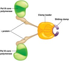
Replication Machinery: The Replisome and Sliding Clamp
Replisome: Large protein complex at the replication fork, coordinating synthesis of both strands.
Sliding clamp: Protein that encircles DNA and tethers DNA polymerase to the template, increasing processivity.
Clamp loader: Loads the sliding clamp onto DNA.
Origins of Replication in Prokaryotes and Eukaryotes
Bacterial origins (e.g., oriC in E. coli): Contain conserved 13-mer and 9-mer sequences recognized by initiator proteins (DnaA, DnaB, DnaC).
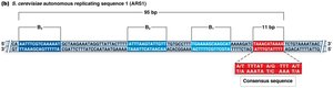
Eukaryotic origins: Multiple origins per chromosome; in yeast, these are called autonomously replicating sequences (ARS).
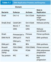
Key Replication Proteins and Enzymes
Domain | Bacteria | Eukarya | Archaea | Role in Replication |
|---|---|---|---|---|
Replication-origin recognition | DnaA | Orc1–6/Cdt1 | Orc1/Cdc6 | Origin recognition |
Helicase activity | DnaB, DnaC | Mcm2–7 | Mcm2–7 | Helicase recruitment |
Primer synthesis | Primase | Primase/pol α | Primase | Primer synthesis |
DNA synthesis | DNA pol III | Pol δ, Pol ε | Pol B, Pol D | DNA synthesis |
Primer removal | DNA pol I, RNaseH | FEN1/DNA2, RNaseH | FEN1/RNaseH | Primer removal |
Sliding clamp | β (sliding) clamp | PCNA | PCNA | DNA polymerase processivity |
Tau protein | τ protein | RFC | RFC | Replication fork progression |
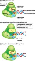
Proofreading and Fidelity of DNA Replication
Proofreading: DNA polymerases possess 3'→5' exonuclease activity, allowing them to remove incorrectly paired nucleotides and replace them with the correct ones.
Accuracy: Replication errors occur at a rate of about one per billion nucleotides in E. coli, largely due to proofreading.
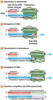
End Replication Problem and Telomeres
Lagging strand issue: The requirement for RNA primers means the very ends of linear chromosomes cannot be fully replicated, leading to progressive shortening.
Telomeres: Repetitive, non-coding sequences at chromosome ends that protect coding DNA from loss.
Telomerase: Ribonucleoprotein enzyme that extends telomeres using an RNA template, active in germ-line and some stem cells.
Clinical Relevance: Telomerase and Disease
Telomerase deficiency: Leads to disorders such as dyskeratosis congenita, characterized by premature aging and stem cell failure.
Telomerase reactivation: Common in cancer cells, allowing unlimited proliferation.
Practice Problems
Draw and label a replication fork, indicating the leading and lagging strands, Okazaki fragments, RNA primers, DNA polymerases, helicase, topoisomerase, primase, SSB, and ligase.
Given the sequence 5’–ATATGCCAAATTCCGG–3’, write the complementary strand in the 5’→3’ direction, and calculate the number of hydrogen and phosphodiester bonds.
Summary
DNA replication is semiconservative and bidirectional, ensuring faithful duplication of the genome.
Multiple enzymes and proteins coordinate to unwind DNA, synthesize new strands, remove primers, and proofread the new DNA.
Telomeres and telomerase are essential for maintaining chromosome integrity in eukaryotes.
