Back
Week 6
Study Guide - Smart Notes
 Tailored notes based on your materials, expanded with key definitions, examples, and context.
DNA Replication, Recombination, and Organization
Overview and Learning Goals
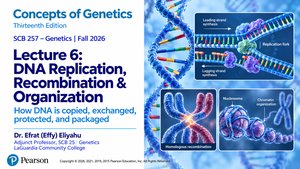
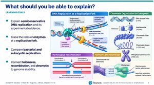
This chapter explores the molecular mechanisms by which DNA is copied, exchanged, protected, and packaged in cells. Key learning objectives include understanding semiconservative DNA replication, the roles of enzymes at the replication fork, comparing bacterial and eukaryotic replication, and connecting telomeres, recombination, and chromatin to genome stability.
Why DNA Replication Is Necessary
Fundamental Requirements
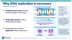
DNA replication is essential for cell division and genetic continuity. The process relies on complementary base pairing, antiparallel strand orientation, and the need to duplicate entire genomes before cell division.
Complementary bases: Each old strand guides the synthesis of a new strand via base pairing (A-T, G-C).
Antiparallel strands: DNA strands run in opposite directions, creating a directionality problem for synthesis.
Genome duplication: Ensures that each daughter cell receives a complete set of genetic information.
Example: Homologous DNA replication produces two identical chromosomes for cell division.
Semiconservative Replication
Central Principle
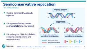
Semiconservative replication describes how each parental DNA strand serves as a template for a new strand, resulting in daughter DNA molecules that each contain one old and one new strand.
Template mechanism: Parental strands separate and guide synthesis of new complementary strands.
Result: Each new DNA double helix contains one parental and one newly synthesized strand.
Models of DNA Replication
Conservative, Semiconservative, and Dispersive Models
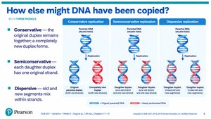
Three models were proposed for DNA replication: conservative, semiconservative, and dispersive. Experimental evidence supports the semiconservative model.
Conservative: Original duplex remains together; new duplex forms separately.
Semiconservative: Each daughter duplex has one original strand.
Dispersive: Old and new segments mix within strands.
Meselson-Stahl Experiment: Key Evidence
Classic Experiment
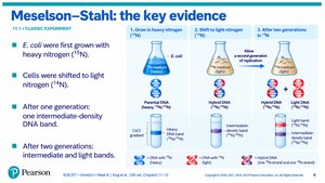
The Meselson-Stahl experiment demonstrated semiconservative replication using isotopic labeling of DNA in E. coli.
Heavy nitrogen (15N): Used to label parental DNA.
Light nitrogen (14N): Shifted cells to new medium; observed intermediate and light DNA bands after generations.
Result: DNA bands confirmed semiconservative replication.
Initiation of Replication
Origins and Replication Bubbles
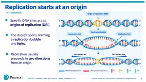
Replication begins at specific DNA sites called origins of replication (ORI). The DNA duplex opens, forming a replication bubble and forks, and replication proceeds in two directions.
Origin of replication: Specific sequence where replication starts.
Replication bubble: Region where DNA is unwound and synthesis begins.
Bidirectional replication: Forks move outward from the origin.
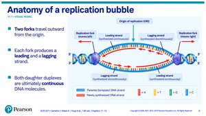
Anatomy of a Replication Bubble
Forks and Strands
Two replication forks travel outward from the origin, each producing a leading and a lagging strand. Both daughter duplexes are ultimately continuous DNA molecules.
Leading strand: Synthesized continuously.
Lagging strand: Synthesized in short fragments (Okazaki fragments).
Replication Toolkit: Enzymes
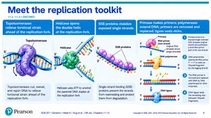
Key Enzymes and Their Functions
Several enzymes coordinate DNA replication, each with a specific role in unwinding, stabilizing, and synthesizing DNA.
Topoisomerase: Relieves torsional strain ahead of the fork.
Helicase: Opens the double helix at the replication fork.
SSB proteins: Stabilize exposed single strands.
Primase: Makes primers; polymerases extend DNA; ligase seals nicks.
Helicase and Single-Strand Binding Proteins
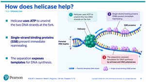
DNA Unwinding and Stabilization
Helicase uses ATP to unwind DNA strands at the fork, while single-strand binding proteins (SSB) prevent immediate reannealing and expose templates for DNA synthesis.
Helicase: Unwinds DNA using ATP.
SSB proteins: Stabilize single-stranded DNA.
Topoisomerases: Preventing DNA Tangling
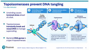
DNA Topology Management
Topoisomerases relieve torsional stress caused by unwinding and transiently break and reseal DNA to manage supercoiling. Bacterial DNA gyrase is a type II topoisomerase.
Unwinding: Creates positive supercoiling ahead of the fork.
Topoisomerase: Relieves supercoiling by breaking and resealing DNA.
DNA gyrase: Specialized for bacterial chromosomes.
DNA Polymerase: Strict Requirements
Core Chemistry
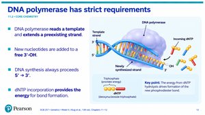
DNA polymerase reads a template and extends a preexisting strand, always adding new nucleotides to a free 3'-OH and synthesizing DNA in the 5' to 3' direction. dNTP incorporation provides the energy for bond formation.
Template reading: Polymerase extends from a preexisting strand.
5' to 3' synthesis: Directionality of DNA synthesis.
Energy: dNTP hydrolysis drives bond formation.
Equation:
Primers in DNA Replication
Why Primers Are Needed
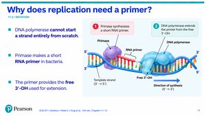
DNA polymerase cannot start a strand from scratch. Primase synthesizes a short RNA primer, which provides the free 3'-OH required for extension.
Primase: Makes RNA primers in bacteria.
3'-OH: Required for DNA polymerase extension.
Antiparallel Strands: The Central Puzzle
Orientation and Synthesis
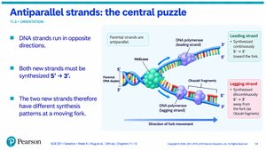
DNA strands run in opposite directions, but both must be synthesized 5' to 3'. This results in different synthesis patterns for the leading and lagging strands at the moving fork.
Antiparallel: Strands run in opposite directions.
Synthesis: Both strands synthesized 5' to 3'.
Products at Each Fork
Leading and Lagging Strands
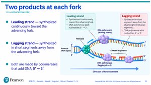
The leading strand is synthesized continuously toward the fork, while the lagging strand is synthesized in short segments away from the fork. Both are made by polymerases that add DNA 5' to 3'.
Leading strand: Continuous synthesis.
Lagging strand: Discontinuous synthesis (Okazaki fragments).
Leading-Strand Synthesis
Continuous Copying
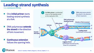
One initial primer starts leading-strand synthesis at a fork. DNA polymerase extends the strand in the direction of fork movement, with continuous extension following the opening helix.
Primer: Starts synthesis.
Continuous extension: Follows fork movement.
Lagging-Strand Synthesis
Discontinuous Copying
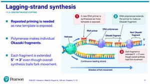
Repeated priming is needed as new template is exposed. Polymerase makes individual Okazaki fragments, each extended 5' to 3' even though overall synthesis trails fork movement.
Repeated priming: Required for lagging strand.
Okazaki fragments: Short DNA segments.
Okazaki Fragment Maturation
Processing Lagging Strand
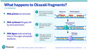
RNA primers are removed, DNA synthesis fills gaps left by removed primers, and DNA ligase seals remaining nicks in the sugar-phosphate backbone.
Primer removal: By RNase H and DNA polymerase.
Gap filling: DNA polymerase fills gaps.
Ligase: Seals nicks.
Leading vs. Lagging: Comparison
Key Differences and Similarities
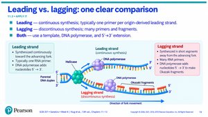
Leading strand synthesis is continuous, typically with one primer per origin-derived strand. Lagging strand synthesis is discontinuous, requiring many primers and fragments. Both use a template, DNA polymerase, and 5' to 3' extension.
Leading: Continuous, one primer.
Lagging: Discontinuous, many primers/fragments.
Both: Template, polymerase, 5' to 3' extension.
Simultaneous Strand Copying
Coordination at the Fork
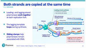
Leading and lagging-strand polymerases work together at each replication fork. The lagging template loops during synthesis, and sliding clamps help polymerases remain associated with DNA.
Coordination: Both strands copied at the same time.
Sliding clamps: Maintain polymerase association.
Proofreading and Replication Accuracy
Genome Integrity
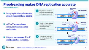
Many replicative polymerases detect incorrect base pairing. A 3' to 5' exonuclease removes misincorporated nucleotides, and polymerase resumes 5' to 3' synthesis after correction.
Proofreading: Detects and corrects errors.
Exonuclease activity: Removes incorrect nucleotides.
Resumption: Synthesis continues after correction.
Replication Fork Checkpoint
Practice Questions
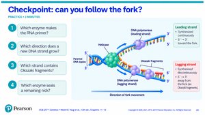
Key questions to test understanding of replication fork mechanics:
Which enzyme makes the RNA primer?
Which direction does a new DNA strand grow?
Which strand contains Okazaki fragments?
Which enzyme seals a remaining nick?
Bacteria vs. Eukaryotes: Replication Logic
Comparative Systems
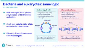
Both bacteria and eukaryotes use origins, forks, primers, polymerases, and bidirectional replication. E. coli uses a single major origin on its circular chromosome, while eukaryotic linear chromosomes have many origins.
Bacteria: Single origin, circular chromosome.
Eukaryotes: Multiple origins, linear chromosomes.
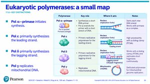
Eukaryotic Polymerases: Key Distinctions
Polymerase Roles and Locations
Eukaryotic cells use several DNA polymerases, each with a distinct role in replication.
Polymerase | Key Role | Where it acts | Notes |
|---|---|---|---|
Pol α (primase) | Initiates synthesis | Replication fork | Starts with RNA primer |
Pol ε | Synthesizes leading strand | Replication fork | High processivity |
Pol δ | Synthesizes lagging strand | Replication fork | Okazaki fragment synthesis |
Pol γ | Replicates mitochondrial DNA | Mitochondria | Specialized for organelles |
Replication and Nucleosomes
Chromatin Challenge
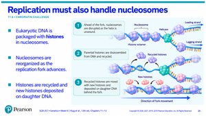
Eukaryotic DNA is packaged with histones in nucleosomes. Nucleosomes are reorganized as the replication fork advances, and histones are recycled and new histones deposited on daughter DNA.
Histones: Package DNA in nucleosomes.
Nucleosome reorganization: Occurs during replication.
Histone recycling: Parental histones mixed with new histones on daughter DNA.