 Back
BackDNA Structure and Analysis: Foundations of Genetic Material
Study Guide - Smart Notes
 Tailored notes based on your materials, expanded with key definitions, examples, and context.
DNA Structure and Analysis
Introduction
This chapter explores the experimental evidence and molecular details that established DNA as the hereditary material. It covers the chemical structure of DNA and RNA, the historical experiments that identified DNA as the genetic material, and the key features of nucleic acid structure.
Characteristics of Hereditary Material
Essential Properties
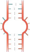
Replication: The genetic material must be able to make accurate copies of itself.
Information Storage: It must store all the information necessary for the structure and function of an organism.
Expression: The information must be expressed to direct cellular processes.
Transmission: It must be passed from parent to offspring.
Mutation: It must be capable of undergoing changes to allow evolution.
Historical Experiments Identifying DNA as Genetic Material
Griffith's Transformation Experiment
Frederick Griffith demonstrated that a 'transforming principle' from dead virulent bacteria could convert non-virulent bacteria into a virulent form, suggesting the transfer of genetic material.
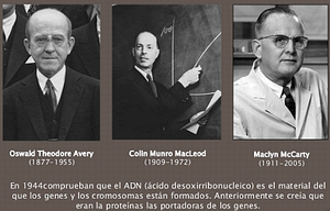
Avery, MacLeod, and McCarty Experiment
These researchers fractionated bacterial extracts and showed that only the DNA fraction could transform non-virulent bacteria, providing strong evidence that DNA is the genetic material.
Hershey-Chase Blender Experiment
Using bacteriophages labeled with radioactive isotopes, Hershey and Chase demonstrated that DNA, not protein, enters bacterial cells and directs viral replication, confirming DNA as the genetic material.
Additional Evidence for DNA as Genetic Material
Organelle Genetics: DNA is present in mitochondria and chloroplasts, which perform genetic functions.
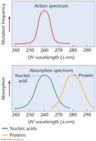
Mutagenesis: DNA and RNA absorb UV light at 260 nm, the same wavelength that induces mutations, while proteins absorb at 280 nm.
Chemical Structure of DNA and RNA
Nucleotides: The Building Blocks
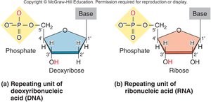
Nucleic acids are polymers of nucleotides, each consisting of a phosphate group, a five-carbon sugar (deoxyribose in DNA, ribose in RNA), and a nitrogenous base.
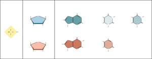
Nitrogenous Bases
Purines: Adenine (A) and Guanine (G) – double-ring structures.
Pyrimidines: Cytosine (C), Thymine (T, in DNA), and Uracil (U, in RNA) – single-ring structures.
Phosphodiester Bonds
Nucleotides are linked by phosphodiester bonds between the 5' phosphate of one nucleotide and the 3' hydroxyl of the next, giving the strand directionality (5' to 3').
Key Experiments Leading to the DNA Double Helix Model
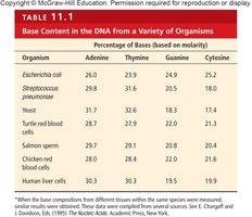
Chargaff's Rules
Erwin Chargaff found that in DNA, the amount of adenine equals thymine (A = T) and the amount of guanine equals cytosine (G = C), suggesting base pairing.
Organism | Adenine (%) | Thymine (%) | Guanine (%) | Cytosine (%) |
|---|---|---|---|---|
Escherichia coli | 26.0 | 23.9 | 24.9 | 25.2 |
Streptococcus | 29.8 | 31.6 | 20.5 | 18.0 |
Yeast | 31.7 | 32.6 | 18.3 | 17.4 |
Turtle red blood cells | 28.0 | 27.9 | 22.0 | 21.3 |
Salmon sperm | 29.2 | 29.2 | 20.8 | 20.4 |
Chicken red blood cells | 28.0 | 28.4 | 21.6 | 21.0 |
Human liver cells | 30.3 | 30.3 | 19.5 | 19.9 |
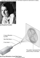
Rosalind Franklin's X-ray Diffraction
Franklin's X-ray diffraction images revealed that DNA is a helical molecule with a regular structure, about 2 nm in diameter, and 10 base pairs per turn.
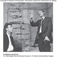
Watson and Crick Model
Watson and Crick integrated Chargaff's rules and Franklin's data to propose the double helix model of DNA, with two antiparallel strands held together by specific base pairing (A-T and G-C).
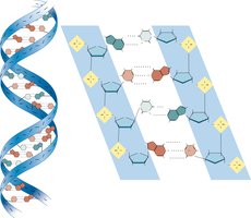
Key Features of the DNA Double Helix
Double Helix: Two strands form a right-handed helix.
Complementary Base Pairing: A pairs with T (2 hydrogen bonds), G pairs with C (3 hydrogen bonds).
Antiparallel Strands: The two strands run in opposite directions (5' to 3' and 3' to 5').
Major and Minor Grooves: The helix has alternating wide (major) and narrow (minor) grooves important for protein binding.
10 Base Pairs per Turn: Each complete turn of the helix contains 10 base pairs.
DNA and RNA: Similarities and Differences
Comparison Table
Feature | DNA | RNA |
|---|---|---|
Sugar | Deoxyribose | Ribose |
Bases | A, T, G, C | A, U, G, C |
Strandedness | Double-stranded (usually) | Single-stranded (usually) |
Function | Genetic information storage | Information transfer, catalysis |
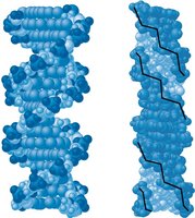
Structural Forms of DNA
B-DNA and Z-DNA
B-DNA: The most common form in cells; right-handed helix, bases perpendicular to axis.
Z-DNA: Left-handed helix, zigzag backbone, can form under certain conditions.
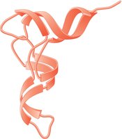
RNA Structure and Function
Primary and Secondary Structure
RNA is typically single-stranded but can form secondary structures such as hairpins, bulges, and internal loops due to complementary base pairing (A-U, C-G).
Major Classes of RNA
mRNA (messenger RNA): Carries genetic information from DNA to ribosomes for protein synthesis.
tRNA (transfer RNA): Brings amino acids to the ribosome during translation; has a cloverleaf secondary structure and L-shaped tertiary structure.
rRNA (ribosomal RNA): Structural and catalytic component of ribosomes.
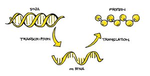
The Central Dogma of Molecular Biology
The central dogma describes the flow of genetic information: DNA is transcribed into RNA, which is then translated into protein.
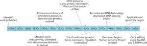
Summary Table: Key Experiments and Discoveries
Year | Discovery |
|---|---|
1860s | Mendel's work published |
1900s | Chromosome theory of inheritance proposed |
1940s | DNA shown to carry genetic information |
1950s | Watson-Crick model of DNA |
1970s | Recombinant DNA technology developed |
1990s | Genomics begins, Human Genome Project initiated |
2000s | Applications of genomics, gene editing technologies |
Key Terms
Virulent: Extremely poisonous or deadly.
Transforming Principle: Substance capable of transferring genetic information.
Mutagenesis: The process of generating mutations.
