Back
BackCh 9 P2 DNA Structure and Analysis – Study Notes
Study Guide - Smart Notes
 Tailored notes based on your materials, expanded with key definitions, examples, and context.
Ch 9 P2 DNA Structure and Analysis
X-Ray Diffraction Analysis
X-ray diffraction analysis is a critical technique used to determine the three-dimensional structure of DNA. Rosalind Franklin's pioneering work in the early 1950s provided key evidence for the helical structure of DNA by analyzing the patterns produced when X-rays are scattered by DNA fibers.
X-ray diffraction involves bombarding DNA molecules with X-rays and capturing the scatter pattern.
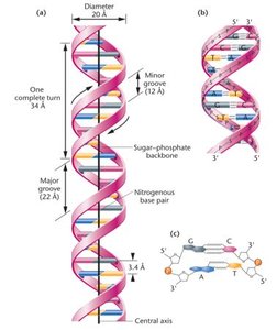
The resulting pattern reveals periodicities and structural features, such as the 3.4 Å spacing between nitrogenous bases, characteristic of a helical structure.
The cross-shaped pattern in the diffraction image indicates the helical nature of DNA.
Example: Franklin's X-ray diffraction images were instrumental in deducing the double helix model of DNA.
The Watson–Crick Model of DNA
James Watson and Francis Crick proposed the double helix model of DNA in 1953, integrating chemical and physical data to describe the molecular structure of DNA.
DNA consists of two antiparallel strands forming a right-handed double helix.
The sugar–phosphate backbones are on the outside, with paired nitrogenous bases on the inside.
There are 10 base pairs per complete turn of the helix, with major and minor grooves present.
The two strands are held together by hydrogen bonds between complementary bases (A-T and G-C).
Example: The antiparallel orientation means one strand runs 5' to 3', while the other runs 3' to 5'.
Chemical Affinity and Complementarity
The specificity of base pairing in DNA is due to chemical complementarity, primarily through hydrogen bonding.
Complementarity refers to the specific pairing of bases: adenine (A) pairs with thymine (T) via two hydrogen bonds, and guanine (G) pairs with cytosine (C) via three hydrogen bonds.
Hydrogen bonds are weak interactions between a covalently bonded hydrogen atom and an electronegative atom with an unshared electron pair.
Example: The higher the G-C content, the more stable the DNA molecule due to the triple hydrogen bonds.
RNA—Ribonucleic Acid
RNA is a nucleic acid similar to DNA but with distinct structural and functional differences.
RNA contains the sugar ribose instead of deoxyribose.
Uracil (U) replaces thymine (T) as a nitrogenous base.
Most RNA molecules are single-stranded, though some viruses have double-stranded RNA.
Example: Messenger RNA (mRNA) is single-stranded and carries genetic information from DNA to ribosomes.
Major Classes of RNA
There are three primary classes of cellular RNA, each playing a distinct role in gene expression.
Messenger RNA (mRNA): Serves as a template for protein synthesis, carrying genetic information from DNA to ribosomes.
Ribosomal RNA (rRNA): Structural and functional component of ribosomes, essential for protein synthesis during translation.
Transfer RNA (tRNA): Delivers amino acids to the ribosome for incorporation into a growing polypeptide chain.
Example: All three RNA types originate as complementary copies of DNA sequences.
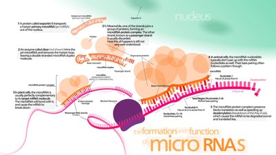
Unique RNAs
In addition to the major classes, several unique RNAs perform specialized functions in the cell.
Telomerase RNA and RNA primers: Involved in DNA replication at chromosome ends (telomeres).
Small nuclear RNA (snRNA): Processes mRNA molecules.
Regulatory RNAs: Includes antisense RNA, microRNA (miRNA), small interfering RNA (siRNA), and long noncoding RNA (lncRNA), all involved in gene regulation.
Analytical Techniques for DNA and RNA
Several analytical techniques are used to study the structure and properties of nucleic acids.
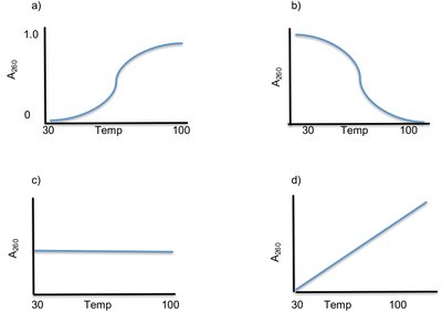
Absorption of UV light (Hyperchromic shift): DNA absorbs UV light maximally at 260 nm. Denaturation increases absorption (hyperchromic effect).
Molecular hybridization: Denatured single strands from different sources can hybridize if complementary.
Fluorescent in situ hybridization (FISH): Uses fluorescent probes to detect specific DNA sequences on chromosomes.
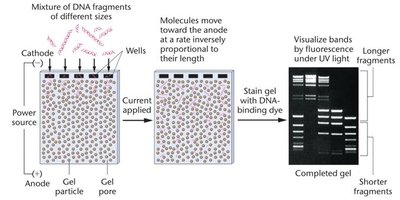
Electrophoresis: Separates DNA and RNA fragments by size using a gel matrix.
Hyperchromic Shift
The hyperchromic shift is observed when double-stranded DNA is heated, causing the strands to separate (denature), resulting in increased UV absorption.
The melting temperature (Tm) is the temperature at which half of the DNA molecules are denatured.
Tm is used to estimate the base composition of DNA; higher G-C content leads to a higher Tm.
Molecular Hybridization
Molecular hybridization involves the denaturation and renaturation of nucleic acids, allowing for the formation of duplex structures between complementary strands from different sources.
DNA strands from different organisms can hybridize if complementary.
RNA can hybridize with the DNA segment from which it was transcribed.
Probes are used to identify specific sequences by hybridization.
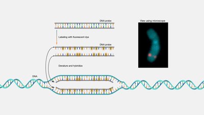
Fluorescent in situ Hybridization (FISH)
FISH is a technique that uses fluorescently labeled probes to detect specific DNA sequences on chromosomes in situ (within the cell).
Mitotic cells are fixed to slides and hybridized with labeled probes.
Probes hybridize only with specific chromosomal regions, allowing visualization under a fluorescence microscope.
Electrophoresis of Nucleic Acids
Electrophoresis is a method used to separate DNA and RNA fragments based on size by applying an electric field to a gel matrix.
Smaller fragments migrate faster through the gel than larger ones.
Agarose gel is commonly used, and DNA bands are visualized using fluorescent dyes.