 Back
BackDNA Structure and Replication: Mechanisms and Experimental Evidence
Study Guide - Smart Notes
 Tailored notes based on your materials, expanded with key definitions, examples, and context.
DNA Structure and Replication
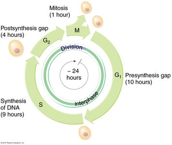
Overview of the Cell Cycle and DNA Replication Timing
The cell cycle is divided into several phases, with DNA replication occurring during the S (synthesis) phase. Accurate duplication of genetic material is essential for cell division and inheritance.
G1 Phase: Presynthesis gap where the cell grows and prepares for DNA replication.
S Phase: DNA synthesis occurs, resulting in chromosome duplication.
G2 Phase: Post-synthesis gap, preparation for mitosis.
M Phase: Mitosis, where chromosomes are segregated into daughter cells.
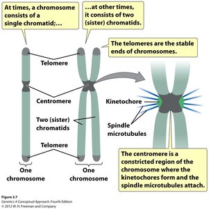
Chromosome Structure and Sister Chromatids
After DNA replication, each chromosome consists of two identical sister chromatids, which are joined at the centromere. These chromatids carry identical alleles of each gene.
Telomeres: Protective structures at the ends of chromosomes.
Centromere: Constricted region where spindle fibers attach during cell division.
Kinetochore: Protein complex at the centromere for microtubule attachment.
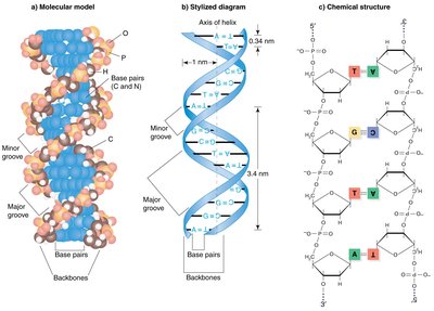
Discovery of DNA Structure
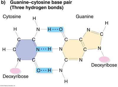
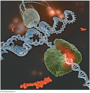
Watson and Crick, using data from Chargaff and Franklin, proposed the double helix model of DNA in 1953. DNA consists of two antiparallel strands forming a right-handed helix, with a sugar-phosphate backbone on the outside and complementary base pairs on the inside.
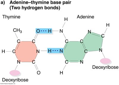
Base Pairing: Adenine (A) pairs with Thymine (T) via two hydrogen bonds; Guanine (G) pairs with Cytosine (C) via three hydrogen bonds.
Antiparallel Strands: One strand runs 5' to 3', the other 3' to 5'.
Properties of Genetic Material
Genetic material must replicate accurately, store information, direct cellular processes, and allow for heritable variation. DNA fulfills all these requirements.
Replication: Ensures progeny inherit identical genetic information.
Information Storage: Encodes instructions for cellular structure and function.
Variation: Mutations introduce heritable changes.
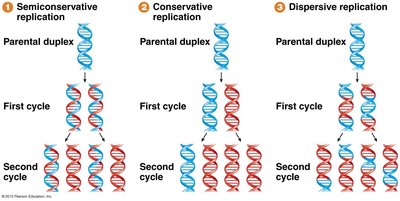
Models of DNA Replication
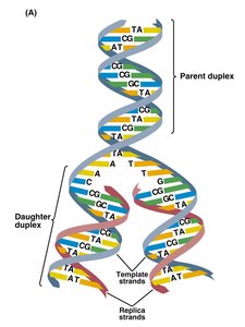
Three models were proposed for DNA replication: semiconservative, conservative, and dispersive. The semiconservative model, supported by experimental evidence, states that each daughter DNA molecule consists of one parental and one newly synthesized strand.
Semiconservative: Each new DNA has one old and one new strand.
Conservative: Parental DNA remains intact; new DNA is entirely new.
Dispersive: Parental and new DNA are interspersed in both strands.
Experimental Evidence: The Meselson-Stahl Experiment
The Meselson-Stahl experiment (1958) used isotopic labeling to distinguish old and new DNA strands, providing strong evidence for the semiconservative model of replication.
Method: E. coli were grown in heavy nitrogen (15N), then transferred to light nitrogen (14N). DNA was extracted after each generation and analyzed by density gradient centrifugation.
Results: After one generation, DNA was of intermediate density (hybrid), ruling out the conservative model. After two generations, both hybrid and light DNA were present, ruling out the dispersive model.


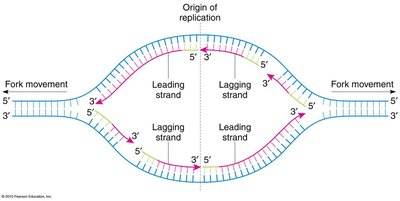
Mechanism of DNA Replication
DNA replication is a highly coordinated process involving multiple enzymes and proteins. Replication begins at origins of replication and proceeds bidirectionally, forming replication forks.
Leading Strand: Synthesized continuously in the direction of fork movement.
Lagging Strand: Synthesized discontinuously in short fragments (Okazaki fragments) opposite to fork movement.
Enzymes Involved: DNA polymerase, helicase, primase, ligase, and single-stranded binding proteins.
DNA Polymerases and Proofreading
DNA polymerases synthesize new DNA by adding nucleotides to a primer strand, following base-pairing rules. They also possess proofreading activity to correct errors.
5' to 3' Synthesis: DNA polymerases add nucleotides only to the 3' end of a growing strand.
3' to 5' Exonuclease Activity: Proofreading function that removes incorrectly paired bases.
Primer Requirement: DNA polymerases require a short RNA primer to initiate synthesis.
Prokaryotic vs. Eukaryotic DNA Replication
Replication in prokaryotes (e.g., E. coli) starts at a single origin of replication, while eukaryotic chromosomes have multiple origins. Eukaryotic chromosomes are linear and contain telomeres at their ends.
Prokaryotes: Single, circular chromosome; one origin of replication.
Eukaryotes: Multiple, linear chromosomes; many origins of replication; presence of telomeres.
Telomeres and Telomerase
Telomeres are repetitive DNA sequences at the ends of linear chromosomes that protect against loss of genetic information during replication. The enzyme telomerase extends telomeres, using an RNA template to add repeats.
Function: Prevents chromosome shortening and loss of essential genes.
Telomerase: A ribonucleoprotein enzyme that adds telomeric repeats to chromosome ends.
