 Back
DNA Structure and Replication: Study Notes for Genetics Students
Study Guide - Smart Notes
 Tailored notes based on your materials, expanded with key definitions, examples, and context.
DNA Structure and Replication
DNA as the Hereditary Molecule
DNA is the fundamental hereditary molecule in all living organisms, responsible for storing and transmitting genetic information. Its identification as the hereditary material was based on several essential characteristics:
Localization: DNA is found in the nucleus and is a component of chromosomes.
Stability: DNA exists in a stable form within cells.
Complexity: DNA contains sufficient information for organismal structure, function, development, and reproduction.
Replication: DNA can accurately replicate itself, ensuring daughter cells inherit identical information.
Mutability: DNA undergoes mutations at a low rate, introducing genetic variation and enabling evolutionary change.
Historical Experiments Identifying DNA as Hereditary Material
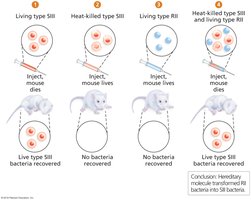
Griffith's Transformation Experiment: Demonstrated that a 'transformation factor' could transfer hereditary traits between bacterial strains.
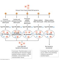
Avery, MacLeod, and McCarty: Showed that DNA, not protein or RNA, is the transformation factor responsible for heredity.
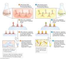
Hershey and Chase: Used radioactive labeling to confirm that DNA, not protein, is the genetic material in bacteriophages.
The DNA Double Helix
The secondary structure of DNA, elucidated by Franklin, Watson, and Crick, consists of two complementary and antiparallel strands forming a double helix. DNA is composed of four nucleotides joined by covalent phosphodiester bonds.
DNA Nucleotides
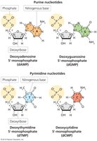
Components: Each nucleotide contains a deoxyribose sugar, a nitrogenous base (adenine, thymine, cytosine, or guanine), and up to three phosphate groups.
Base Types:
Pyrimidines: Thymine (T) and cytosine (C) – single ring.
Purines: Adenine (A) and guanine (G) – double ring.
Polynucleotide Chain Formation
Enzyme: DNA polymerase catalyzes the formation of phosphodiester bonds between the 3' hydroxyl group of one nucleotide and the 5' phosphate of another.
Backbone: Alternating sugar and phosphate groups form the backbone of each strand.
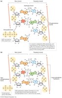
Complementary and Antiparallel Strands
Complementarity: A pairs with T (2 hydrogen bonds), G pairs with C (3 hydrogen bonds).
Antiparallel: The two strands run in opposite directions (5' to 3' and 3' to 5').
Structural Features of the Double Helix
Base Pair Spacing: Nucleotide base pairs are spaced at intervals of 3.4 Å.
Helix Diameter: B-form DNA has a diameter of 20 Å.
Base Stacking: Tight packing and parallel planes of base pairs lead to the helical twist.
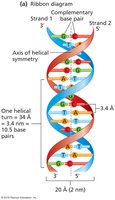
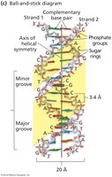
Major and Minor Grooves
Major Groove: ~12 Å wide; allows protein binding.
Minor Groove: ~6 Å wide; also accessible to proteins.
Forms of DNA
B-form: Most common, right-handed helix.
A-form: Right-handed, found in some cellular contexts.
Z-form: Left-handed, zigzag backbone, often near transcription start sites.
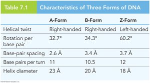
Form | Helical Twist | Rotation per Base Pair | Base-Pair Spacing | Base Pairs per Turn | Helix Diameter |
|---|---|---|---|---|---|
A-Form | Right-handed | 32.7° | 2.6 Å | 11 | 23 Å |
B-Form | Right-handed | 34.3° | 3.4 Å | 10.5 | 20 Å |
Z-Form | Left-handed | 60.2° | 3.7 Å | 12 | 18 Å |
DNA Replication: Mechanisms and Models
DNA replication is essential for genetic continuity and occurs with high fidelity. The process is semiconservative and bidirectional in most organisms.
Attributes of DNA Replication
Each parental strand remains intact and serves as a template.
Each daughter duplex contains one parental and one daughter strand.
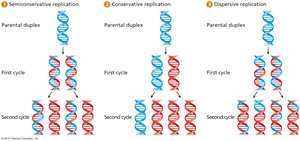
Models of DNA Replication
Semiconservative: Each daughter duplex contains one parental and one daughter strand.
Conservative: One duplex contains both parental strands, the other both daughter strands.
Dispersive: Each duplex contains interspersed parental and daughter segments.
Origin and Directionality of Replication
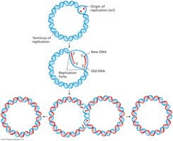
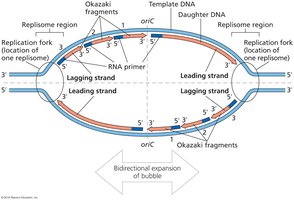
Bacterial Chromosomes: Replication is bidirectional from a single origin (oriC).
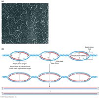
Eukaryotic Chromosomes: Multiple origins of replication exist.
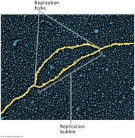
Replication Bubble: Expansion occurs around the origin, with replication forks at each end.
Multiple Replication Origins in Eukaryotes
Electron Micrograph Evidence: Shows multiple origins on chromosomes.
Yeast ARS: Autonomously replicating sequences are well characterized in yeast.
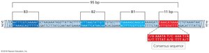
Molecular Biology of Replication Initiation
Replication initiation involves specific proteins binding to origin sequences and the synthesis of RNA primers.
Bacteria: DnaA, DnaB, and DnaC bind to oriC.
Archaea: Orc1/Cdc6 binds to ORB and miniORB sequences.
Eukaryotes: Prereplication complex includes the origin replication complex (ORC).
Primase: Synthesizes RNA primers required for DNA polymerase activity.
Continuous and Discontinuous Strand Replication
DNA polymerase III synthesizes the leading strand continuously and the lagging strand discontinuously via Okazaki fragments.
Leading Strand: Synthesized in the direction of fork progression.
Lagging Strand: Synthesized in short segments (Okazaki fragments) opposite to fork progression.
RNA Primer Removal and Okazaki Fragment Ligation
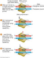
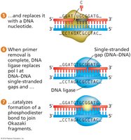
DNA Polymerase I: Removes RNA primers (5'-3' exonuclease) and adds DNA nucleotides (5'-3' polymerase).
DNA Ligase: Seals gaps between DNA segments by forming phosphodiester bonds.
DNA Proofreading
DNA polymerases possess proofreading activity, ensuring high fidelity during replication. Errors are corrected by 3'-5' exonuclease activity.
Error Rate: Approximately one error per billion nucleotides in E. coli.
Mechanism: Incorrect nucleotides are removed and replaced.
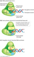
Replication at the Ends of Linear Chromosomes: Telomeres
Telomeres are repetitive sequences at chromosome ends that protect against loss of genetic information during replication. The lagging strand cannot be fully replicated, but telomeres ensure stability.
Telomerase: A ribonucleoprotein enzyme that extends telomeres in germ line and certain cells.
Telomere Shortening: Occurs in most somatic cells, associated with aging and cancer.
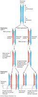
Methods of Molecular Genetic Analysis
DNA replication processes are utilized in molecular genetic techniques such as PCR and DNA sequencing. These methods allow for the amplification and analysis of genetic material.
PCR: Polymerase chain reaction amplifies specific DNA sequences.
VNTR Analysis: Variable number tandem repeat alleles are separated after PCR amplification for genotyping.
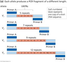
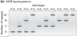
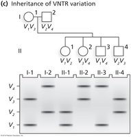
Example: PCR and VNTR Analysis
Each allele produces a PCR fragment of different length based on the number of repeats.
Band patterns can be used to determine genotype and inheritance.
Key Equations and Concepts
Phosphodiester Bond Formation:
Base Pairing:
Semiconservative Replication:
Additional info: The notes expand on brief points with academic context, definitions, and examples to ensure completeness and clarity for Genetics students.
