 Back
BackDNA Structure, Function, and Experimental Evidence: Study Notes for Genetics Students
Study Guide - Smart Notes
 Tailored notes based on your materials, expanded with key definitions, examples, and context.
DNA as Genetic Material
Experimental Evidence for DNA as Genetic Material
The identification of DNA as the genetic material was established through a series of landmark experiments. These experiments demonstrated that DNA, not protein, is responsible for heredity in living organisms.
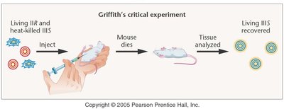
Griffith’s Experiment: Frederick Griffith showed that a 'transforming principle' could transfer genetic information from dead virulent bacteria to live non-virulent bacteria, making them virulent. This suggested that some molecular component was responsible for heredity.
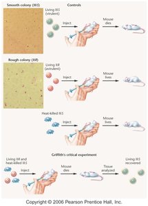
Transformation Principle: The ability of non-virulent bacteria to become virulent after exposure to heat-killed virulent bacteria indicated the presence of a transferable genetic material.
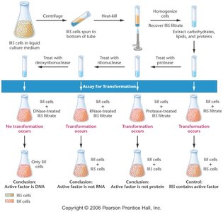
Avery, McLeod, and McCarty’s Experiment: These scientists isolated and treated the transforming principle with enzymes that degrade DNA, RNA, and protein. Only DNA destruction prevented transformation, confirming DNA as the genetic material.
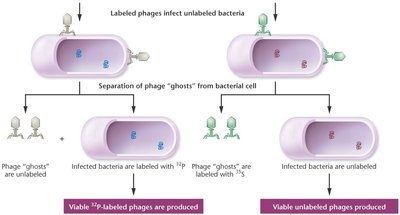
Hershey-Chase Experiment: Using radioactive labeling, Hershey and Chase showed that only DNA, not protein, entered bacteria during phage infection, proving DNA is the genetic material in viruses.
Structural Characteristics of DNA
Nucleic Acid Composition
DNA and RNA are composed of three main components: nitrogenous bases, pentose sugars, and phosphate groups. The arrangement of these components forms the backbone and coding regions of nucleic acids.
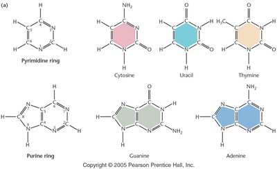
Nitrogenous Bases: There are two types of nitrogenous bases:
Pyrimidines: Cytosine, Thymine (DNA), Uracil (RNA)
Purines: Adenine, Guanine
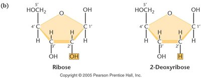
Pentose Sugars: DNA contains 2-deoxyribose, while RNA contains ribose. The difference is the presence or absence of a hydroxyl group at the 2' carbon.
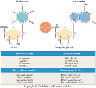
Nucleosides and Nucleotides:
Nucleoside: Nitrogenous base + sugar
Nucleotide: Nucleoside + phosphate group
Nucleoside Triphosphate (NTP): NTPs are the activated forms used in DNA and RNA synthesis, containing three phosphate groups.
Physical Properties of DNA Molecule
DNA molecules exhibit specific physical properties, including double-strandedness, linear or circular structure, and length. These properties vary between viruses, bacteria, and eukaryotes.
Viral DNA Genome: Can be single- or double-stranded, linear or circular, and is inert when packaged.
Bacterial Genome: Typically double-stranded, linear DNA, associated with a few DNA-binding proteins, and not functionally inert.
Genome Organization
Eukaryotic Genome: Chromatin Structure and Nucleosomes
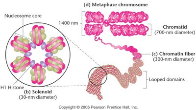
In eukaryotes, DNA is organized into chromatin, which is a complex of DNA and histone proteins. Chromatin structure is essential for DNA packaging, regulation, and accessibility.
Chromatin: DNA uncoils during interphase to form chromatin, which is always associated with histones.
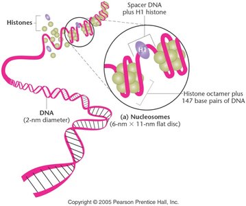
Histones: Basic, positively charged proteins (H1, H2A, H2B, H3, H4) rich in arginine and lysine. The structural unit of chromatin is the nucleosome.
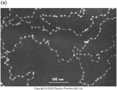
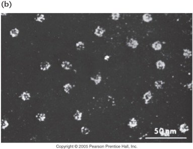
Nucleosome: Consists of a histone octamer (two each of H2A, H2B, H3, H4) wrapped by 147 base pairs of DNA. H1 histone acts as a linker.
Chromatin Remodeling: Histones can be chemically modified, affecting chromatin structure and gene expression.
Techniques Used to Study DNA
Labeling and Visualization
Various techniques are used to study DNA, including radioactive labeling, electron microscopy, and biochemical assays. These methods help elucidate DNA structure, function, and organization.
Radioactive Labeling: Used in experiments such as Hershey-Chase to distinguish DNA from protein.
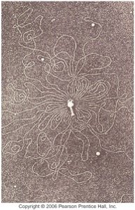
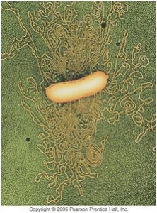
Electron Microscopy: Allows visualization of DNA molecules and chromatin structure.
Biochemical Assays: Used to identify DNA, RNA, and protein components in cells.
Summary Table: Nucleosides and Nucleotides
The following table summarizes the names of nucleosides and nucleotides for both RNA and DNA:
Type | Nucleoside | Nucleotide |
|---|---|---|
RNA | Adenosine, Cytidine, Guanosine, Uridine | Adenylic acid, Cytidylic acid, Guanylic acid, Uridylic acid |
DNA | Deoxyadenosine, Deoxycytidine, Deoxyguanosine, Deoxythymidine | Deoxyadenylic acid, Deoxycytidylic acid, Deoxyguanylic acid, Deoxythymidylic acid |
Key Equations
Phosphodiester Bond Formation: The backbone of DNA is formed by phosphodiester bonds between the 3' hydroxyl group of one nucleotide and the 5' phosphate group of the next.
Conclusion
Understanding the structure, function, and experimental evidence for DNA as the genetic material is foundational in genetics. The organization of DNA into chromatin and nucleosomes, as well as the techniques used to study DNA, are essential concepts for further exploration in molecular genetics.
