 Back
BackDNA Structure: The Molecular Basis of Heredity
Study Guide - Smart Notes
 Tailored notes based on your materials, expanded with key definitions, examples, and context.
DNA Structure and the Molecular Basis of Heredity
Introduction to Hereditary Material
The hereditary material is the molecular substance responsible for carrying and transmitting genetic information from one generation to the next. DNA (deoxyribonucleic acid) is now recognized as the primary hereditary molecule in all cellular life forms.
Definition: The hereditary molecule must be localized to chromosomes, present in a stable form, sufficiently complex, able to replicate accurately, and mutable to allow genetic variation.
Historical context: Early candidates for hereditary material included DNA, RNA, proteins, lipids, and carbohydrates.
Essential Features of Hereditary Material
Localization: Found in the nucleus and associated with chromosomes.
Stability: Maintains a stable presence in cells across generations.
Complexity: Encodes information for structure, function, development, and reproduction.
Replication: Capable of accurate self-replication, ensuring daughter cells inherit identical information.
Mutability: Undergoes rare mutations, providing the basis for genetic diversity and evolution.
Experimental Evidence for DNA as the Hereditary Molecule
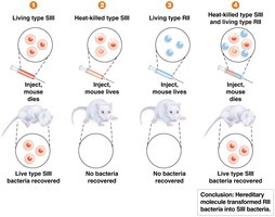
Griffith's Transformation Experiment
Frederick Griffith's experiments with Pneumococcus bacteria demonstrated the existence of a 'transformation factor' capable of transferring hereditary information.
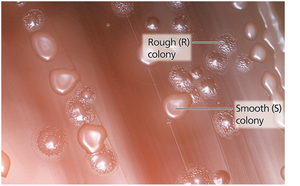
S (smooth) strain: Virulent, causes fatal pneumonia in mice.
R (rough) strain: Non-virulent, does not cause disease.
Key finding: Mixing heat-killed S strain with live R strain transformed the R strain into a virulent form, indicating transfer of genetic material.
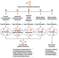
Avery, MacLeod, and McCarty's Experiment
These researchers identified DNA as the 'transformation factor' by selectively destroying different macromolecules in extracts from heat-killed S bacteria.
Method: Treated extracts with enzymes to destroy proteins, RNA, or DNA.
Result: Only destruction of DNA prevented transformation, confirming DNA as the hereditary molecule.
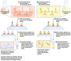
Hershey-Chase Experiment
Alfred Hershey and Martha Chase used bacteriophages labeled with radioactive isotopes to show that DNA, not protein, enters bacterial cells and directs viral replication.
Labeling: DNA labeled with 32P, protein with 35S.
Conclusion: Only DNA entered the bacteria and was required for viral replication.
Discovery and Structure of DNA
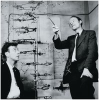
Watson and Crick Model
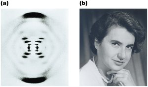
James Watson and Francis Crick, using data from Rosalind Franklin's x-ray diffraction studies and Chargaff's rules, proposed the double helix structure of DNA in 1953.
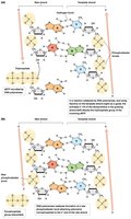
Double helix: Two antiparallel strands with sugar-phosphate backbones on the outside and nitrogenous bases paired in the center.
Complementary base pairing: Adenine pairs with thymine (A-T), guanine pairs with cytosine (G-C).
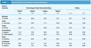
Chargaff's Rule
Erwin Chargaff discovered that the amount of adenine equals thymine and the amount of guanine equals cytosine in DNA from various organisms.
Chargaff's rule: [A] = [T] and [G] = [C]
Implication: Base pairing is specific and consistent across species.
Genome | Adenine (%) | Guanine (%) | Cytosine (%) | Thymine (%) | G/C Ratio |
|---|---|---|---|---|---|
E. coli | 23.8 | 26.0 | 25.3 | 23.9 | 1.02 |
S. cerevisiae | 31.3 | 18.7 | 17.3 | 32.5 | 1.09 |
H. sapiens | 29.8 | 20.7 | 19.8 | 29.9 | 1.05 |
DNA Nucleotide Structure
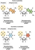
Components of a DNA Nucleotide
Each DNA nucleotide consists of a deoxyribose sugar, a phosphate group, and one of four nitrogenous bases: adenine (A), guanine (G), cytosine (C), or thymine (T).
Deoxyribose: Five-carbon sugar lacking an OH group at the 2' position.
Nitrogenous bases: Purines (A, G) have double rings; pyrimidines (C, T) have single rings.
Phosphate group: Attached to the 5' carbon of the sugar.
Formation of Polynucleotide Chains
Nucleotides are joined by phosphodiester bonds between the 3' hydroxyl of one nucleotide and the 5' phosphate of the next, forming a sugar-phosphate backbone.
Directionality: Each strand has a 5' end (phosphate) and a 3' end (hydroxyl).
Enzyme: DNA polymerase catalyzes the formation of these bonds during DNA synthesis.
Double Helix and Base Pairing
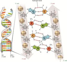
Complementary and Antiparallel Strands
The two DNA strands are complementary, with specific base pairing (A-T, G-C), and run in opposite directions (antiparallel).
Hydrogen bonds: Two between A and T, three between G and C.
Antiparallel orientation: One strand runs 5' to 3', the other 3' to 5'.
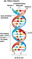
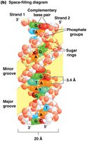
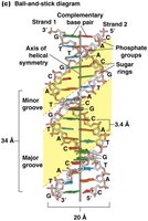
Helical Structure and Dimensions
The DNA double helix has a uniform diameter of 20 Å and completes one turn every 34 Å, with base pairs stacked 3.4 Å apart.
Base stacking: Adjacent base pairs are offset, creating a helical twist.
Major and minor grooves: Alternating grooves provide binding sites for DNA-binding proteins.
Summary Table: Key Features of DNA Structure
Feature | Description |
|---|---|
Hereditary Material | DNA is the primary molecule carrying genetic information |
Double Helix | Two antiparallel strands with complementary base pairing |
Nucleotide Components | Deoxyribose sugar, phosphate group, nitrogenous base |
Base Pairing | A-T (2 H bonds), G-C (3 H bonds) |
Grooves | Major (12 Å) and minor (6 Å) grooves for protein binding |
Additional info: This guide covers the foundational concepts of DNA structure, its role as hereditary material, and the experimental evidence supporting these conclusions. For further study, review the chemical mechanisms of DNA replication and the regulation of gene expression.
