 Back
Eukaryotic Chromosome Abnormalities and Molecular Organization: Study Guide
Study Guide - Smart Notes
 Tailored notes based on your materials, expanded with key definitions, examples, and context.
Eukaryotic Chromosome Abnormalities and Molecular Organization
Chromosome Number and Shape Vary among Organisms
Chromosome number, size, and shape are species-specific and play a fundamental role in heredity and genetic variation. Closely related species tend to have similar chromosome numbers, but the content and structure of chromosomes are unique to each species.
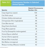
Genome Content: The total chromosome number in a nucleus varies widely among species.
Chromosome Pair Distinction: Each chromosome pair in a diploid genome has a distinct size, shape, and genetic content.
Visualization: Chromosomes can be viewed using various cytogenetic techniques.
Example: Table 10.1 shows diploid chromosome numbers for selected animal species.
Chromosomes in Nuclei
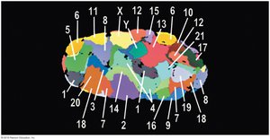
Chromosomes are not uniformly distributed within the nucleus. They occupy specific regions called chromosome territories during interphase, which are important for gene regulation and nuclear organization.
Chromosome Territories: Chromosomes are partitioned into distinct regions within the nucleus.
Dynamic Nature: Chromosomes move, twist, and turn within their territories during transcription and DNA replication.
Anchoring: Chromosomes are anchored by their centromeres.
Interchromosomal Domains: Channels between territories allow movement of proteins, enzymes, and RNA molecules.
Gene-Rich Chromosomes: Larger, gene-rich chromosomes are near the center; smaller, gene-poor chromosomes are near the periphery.
Karyotypes
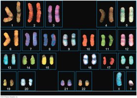
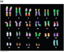
A karyotype is an organized visual display of chromosomes, used to identify abnormalities in chromosome number or structure. Chromosome condensation is maximal at metaphase, and staining techniques help distinguish chromosomes.
Autosomal Homologs: Numbered 1 through 22 by size; sex chromosomes are identified separately.
Staining: Different compounds are used to stain chromosomes for identification.
Chromosome Shape and Karyotypes
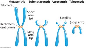
Chromosome size and shape are important for ordering chromosomes in a karyotype. The centromere divides chromosomes into arms of unequal length, and the position of the centromere determines chromosome shape.
Chromosome Arms: Short arm (p), long arm (q).
Centromere Position: Determines chromosome shape: metacentric, submetacentric, acrocentric, telocentric.
Fluorescent In Situ Hybridization (FISH)
FISH uses molecular probes labeled with fluorescent compounds to detect specific DNA sequences. Multiple probes with different fluorescent labels can be used simultaneously to identify gene-specific sequences.
Application: Used for gene mapping and detection of chromosomal abnormalities.
Chromosome Banding Techniques
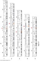
Chromosome banding allows cytogeneticists to identify chromosomes based on size, shape, and banding pattern. Giemsa (G) banding is the standard for human chromosomes.
Banding Patterns: Distinct and reproducible for each chromosome.
Preparation: Includes cell culture, metaphase arrest, and slide preparation.
Heterochromatin and Euchromatin
Chromosome condensation varies along the chromosome. Regions with actively expressed genes are less condensed (euchromatin), while tightly condensed regions with fewer expressed genes are called heterochromatin.
Euchromatin: Less condensed, gene-rich, actively transcribed.
Heterochromatin: Highly condensed, gene-poor, transcriptionally inactive.
Nondisjunction and Chromosome Number Abnormalities
Nondisjunction Leads to Changes in Chromosome Number
Nondisjunction is the failure of chromosomes or sister chromatids to separate properly during cell division, resulting in abnormal chromosome numbers (aneuploidy).
Euploid: Complete sets of chromosomes (e.g., n, 2n, 3n).
Aneuploid: Chromosome number not a complete set.
Nondisjunction in Germ-Line Cells
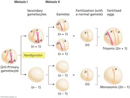
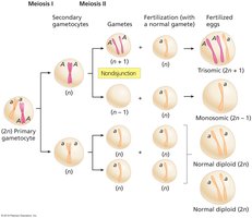
Nondisjunction in meiosis I results in gametes with n+1 or n-1 chromosomes, leading to trisomic (2n+1) or monosomic (2n-1) offspring after fertilization.
Meiosis I: Failure of homologs to separate.
Meiosis II: Failure of sister chromatids to separate; two normal gametes, one n+1, one n-1.
Aneuploidy in Humans
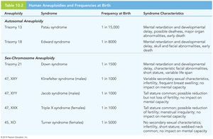
Humans are sensitive to gene dosage changes; most aneuploids do not survive. Only trisomies of chromosomes 13, 18, and 21 are seen in newborns, and no autosomal monosomies are observed. Multiple forms of sex-chromosome aneuploidy occur.
Trisomy 21 (Down syndrome): Associated with cognitive disabilities and heart abnormalities; risk increases with maternal age.
Turner syndrome (XO): Monosomy of X chromosome; haploinsufficiency of SHOX gene causes symptoms.
Mosaicism and Uniparental Disomy
Mosaicism results from mitotic nondisjunction early in embryogenesis, leading to individuals with different cell lines. Uniparental disomy occurs when both copies of a chromosome pair come from the same parent, associated with syndromes like Angelman and Prader-Willi.
Mosaicism: Presence of two or more cell lines with different chromosome numbers.
Uniparental Disomy: Both homologs from one parent; can cause genetic disorders.
Polyploidy and Changes in Euploid Content
Polyploidy
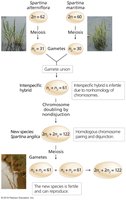
Polyploidy is the presence of three or more sets of chromosomes. It can arise from duplication within a species (autopolyploidy) or from combining chromosome sets of different species (allopolyploidy).
Autopolyploidy: Duplication of chromosome sets within a species.
Allopolyploidy: Combination of chromosome sets from different species.
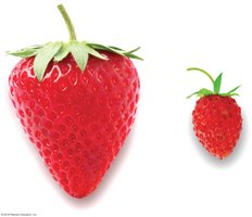
Effects: Increased fruit and flower size, decreased fertility.
Chromosome Breakage and Structural Mutations
Chromosome Breakage Causes Mutation by Loss, Gain, and Rearrangement
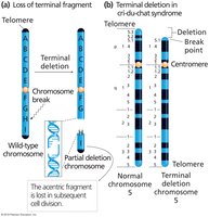
Chromosome breakage can lead to loss or gain of chromosome segments, causing gene dosage imbalances and severe abnormalities.
Partial Deletion: Loss of terminal or internal chromosome segments.
Partial Deletion Heterozygotes: One normal and one deleted chromosome; example: cri-du-chat syndrome.
Interstitial Deletion: Loss of internal chromosome portion due to two breaks.
Unequal Crossover
Unequal crossover between homologs can result in partial duplication on one homolog and partial deletion on the other. This is often due to misalignment of repetitive regions.
Partial Duplication Heterozygotes: One normal and one duplication homolog.
Example: Williams-Beuren syndrome involves partial deletion of chromosome 7.
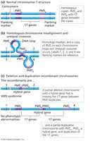
Detecting Duplication and Deletion
Large deletions or duplications can be detected by microscopy, while microdeletions and microduplications are detected using molecular techniques such as FISH.
Microscopy: Reveals altered banding patterns.
FISH: Detects absence or duplication of DNA sequences.
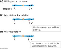
Microscopic Observation of Synapsis
During prophase I, regions of chromosome duplication or deletion can be observed as unpaired loops at synapsis.
Unpaired Loop: Indicates mismatch due to duplication or deletion.
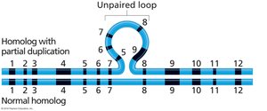
Deletion Mapping
Pseudodominance occurs when a recessive allele is expressed due to deletion of the dominant allele. Deletion mapping is used to pinpoint gene locations by observing pseudodominance in heterozygotes.
Application: Used to map gene positions on chromosomes.
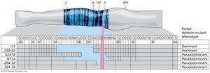
Chromosome Inversion and Translocation
Chromosome Inversion
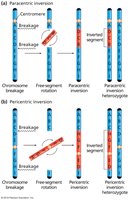
Chromosome breakage and reattachment in the wrong orientation leads to inversion. Two types exist: paracentric (centromere outside inverted region) and pericentric (centromere within inverted region).
Inversion Heterozygotes: One normal and one inverted homolog.
Chromosome Pairing and Recombination
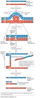
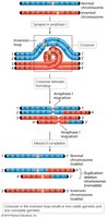
Alignment of normal and inverted homologs forms an inversion loop at synapsis. Crossing over within the inverted region results in duplications and deletions in recombinant chromosomes.
Paracentric Inversion: Crossing over produces dicentric and acentric chromosomes.
Pericentric Inversion: Crossing over produces chromosomes with duplicated and deleted regions.
Chromosome Translocation
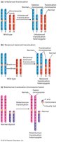
Translocations occur when broken ends of nonhomologous chromosomes are reattached. Translocation heterozygotes may be phenotypically normal but can experience semisterility due to segregation abnormalities.
Types: Nonreciprocal, reciprocal balanced, Robertsonian (chromosome fusion).
Eukaryotic Chromosome Organization into Chromatin
Chromatin Structure and Function
Chromatin organization is essential for chromosome function, distribution during cell division, and regulation of gene expression. Chromosomes are composed of approximately half DNA and half protein, including histones and nonhistone proteins.
Histones: Small, basic proteins that tightly bind DNA; five types: H1, H2A, H2B, H3, H4.
Nucleosome: Core particle with two molecules each of H2A, H2B, H3, and H4; DNA (~146 bp) wraps around octamer.
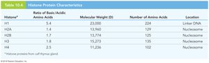
Nucleosome Assembly
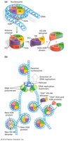
Histones assemble into dimers and tetramers to form the nucleosome octamer. DNA wrapping around the octamer is the first level of condensation, compacting DNA sevenfold.
Assembly: H2A-H2B dimers, H3-H4 dimers, H3-H4 tetramer, octamer formation.
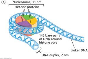
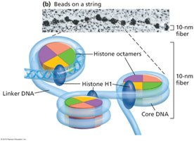
Chromatin Structure: Beads-on-a-String
Electron micrographs show chromatin in its least condensed state as a 10-nm fiber, or "beads-on-a-string" morphology. The beads are nucleosomes, and the string is linker DNA.
Linker DNA: Variable-length DNA between nucleosomes.
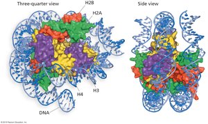
Solenoid Structure and Higher-Order Organization
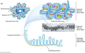
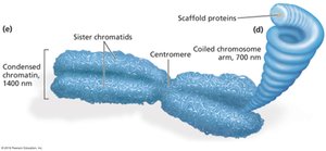
The 10-nm fiber coils into a 30-nm solenoid structure under normal cellular conditions, stabilized by histone H1. Chromatin exists as 30-nm fibers or more condensed states during interphase and becomes maximally condensed during metaphase.
Solenoid: Six to eight nucleosomes per turn.
Chromosome Scaffold: Loops of 30-nm fibers attached to nonhistone protein scaffold.
Nucleosome Disassembly and Reassembly during Replication
During DNA replication, nucleosomes are partially disassembled and reassembled from old and new histone components. Most nucleosomes after replication are hybrids of old and new histones.
Histone Inheritance: Old histones retained as single molecules, dimers, or tetramers.
Position Effect Variegation: Chromatin State and Transcription
Chromatin compaction regulates access to DNA for transcription. Position effect variegation (PEV) in Drosophila demonstrates how chromatin state can control gene expression or silencing, which is transmissible from one cell generation to the next.
Gene Expression: Controlled by chromatin state.
Gene Silencing: Dictated by chromatin structure.
