 Back
Eukaryotic Chromosome Abnormalities and Molecular Organization: Study Notes
Study Guide - Smart Notes
 Tailored notes based on your materials, expanded with key definitions, examples, and context.
Chapter 10: Eukaryotic Chromosome Abnormalities and Molecular Organization
10.1 Chromosome Number and Shape Vary Among Organisms
Chromosome number, size, and shape are species-specific and play a fundamental role in heredity and genetic variation. Chromosomes are distributed non-uniformly within the nucleus, and their organization is crucial for gene expression and cellular function.
Genome Content: The total chromosome number in a nucleus varies widely among species, but closely related species tend to have similar numbers.
Chromosome Territories: Chromosomes occupy distinct regions within the nucleus during interphase, known as chromosome territories.
Chromosome Pairing: Each chromosome pair in a diploid genome has unique size, shape, and genetic content.
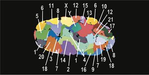
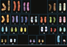
Karyotypes
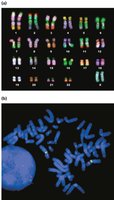
A karyotype is an organized visual display of chromosomes, used to identify abnormalities in chromosome number or structure. Autosomal homologs are numbered 1–22, and sex chromosomes are identified separately. Chromosomes can be stained to distinguish them.
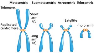
Chromosome Shapes
Chromosomes are classified based on the position of the centromere:
Metacentric: Centromere near the middle.
Submetacentric: Centromere between center and tip.
Acrocentric: Centromere close to one end.
Telocentric: Centromere at the tip, no p arm.
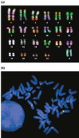
Fluorescent in Situ Hybridization (FISH)
FISH uses fluorescently labeled molecular probes to detect specific DNA sequences on chromosomes. Multiple probes emitting different wavelengths can be used simultaneously to identify various sequences.
Heterochromatin and Euchromatin
Chromosome condensation varies along its length, affecting gene expression:
Euchromatin: Less condensed, actively expressed genes.
Heterochromatin: Highly condensed, fewer expressed genes.
Each G-band contains 1–10 million base pairs and multiple genes.
10.2 Nondisjunction Leads to Changes in Chromosome Number
Chromosome Nondisjunction
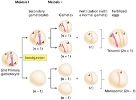
Nondisjunction is the failure of chromosomes or chromatids to separate properly during cell division, resulting in abnormal chromosome numbers (aneuploidy).
Euploid: Complete sets of chromosomes.
Aneuploid: Chromosome number not a complete set.
Nondisjunction in Germ-Line Cells
Nondisjunction during meiosis produces aneuploid gametes, which can lead to aneuploid zygotes. Fusion with normal gametes results in trisomic or monosomic offspring.
Aneuploidy in Humans
Humans are sensitive to gene dosage changes; most aneuploids do not survive. Only trisomies of chromosomes 13, 18, and 21 are seen in newborns, and no autosomal monosomies are observed. Sex chromosome aneuploidies are more common.
Trisomy 21 (Down Syndrome)
Trisomy 21 is linked to maternal age. Prolonged meiotic arrest in oocytes may degrade cohesin proteins, increasing nondisjunction risk. A few genes on chromosome 21 cause the main symptoms.
Turner Syndrome
Turner syndrome (45, XO) results from monosomy of the X chromosome. Haploinsufficiency of the SHOX gene, which escapes X-inactivation, is a primary cause of short stature and developmental abnormalities.
10.3 Changes in Euploid Content Lead to Polyploidy
Polyploidy
Polyploidy is the presence of three or more sets of chromosomes. It can arise from duplication within a species (autopolyploidy) or from combining sets of different species (allopolyploidy).
Polyploidy is common in plants and can lead to new species.
Polyploid organisms often have increased size and vigor.
10.4 Chromosome Breakage Causes Mutation by Loss, Gain, and Rearrangement of Chromosomes
Partial Chromosome Deletion
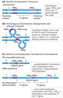
Chromosome breakage at a break point can lead to terminal deletions. Acentric fragments (lacking centromeres) are often lost during cell division.
Unequal Crossover
Unequal crossover between homologs results in partial duplication on one homolog and partial deletion on the other. This can cause syndromes such as Williams-Beuren syndrome.
10.5 Chromosome Breakage Leads to Inversion and Translocation of Chromosomes
Chromosome Inversion
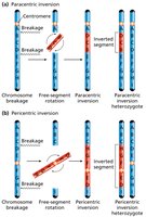
Chromosome breakage and reattachment in the wrong orientation leads to inversion. Two types:
Paracentric inversion: Centromere outside inverted region.
Pericentric inversion: Centromere within inverted region.
Chromosome Pairing and Recombination
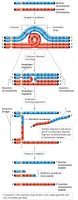
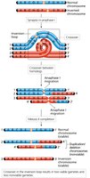
Alignment of normal and inverted homologs forms an inversion loop. Crossing over outside the inverted region is normal, but within the region causes duplications and deletions.
Crossing Over Within a Paracentric Inversion
Results in a dicentric chromosome (pulled to both poles, breaks randomly) and an acentric fragment (lost).
Crossing Over Within a Pericentric Inversion
Results in duplicated and deleted regions in recombinant chromosomes. Two normal and two abnormal gametes are produced.
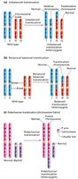
Three Types of Translocations
Nonreciprocal translocation: Piece of one chromosome moves to a nonhomolog.
Reciprocal balanced translocation: Pieces of two nonhomologs switch places.
Robertsonian translocation: Fusion of two nonhomologous chromosomes.
10.6 Eukaryotic Chromosomes Are Organized into Chromatin
Chromatin Organization
Chromatin structure is essential for chromosome function, gene regulation, and compaction within the nucleus.
Chromatin is composed of DNA and proteins, mainly histones.
Histone proteins are highly conserved and critical for nucleosome formation.
Histone Proteins and Nucleosomes
There are five types of histone proteins: H1, H2A, H2B, H3, and H4. Nucleosome core particles consist of an octamer of histones (two each of H2A, H2B, H3, and H4), with DNA wrapped around them.
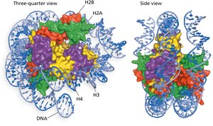
Nucleosome Assembly
Two H3 and two H4 form a tetramer, which binds DNA.
Two H2A-H2B dimers associate to complete the octamer.
Chromatin Structure
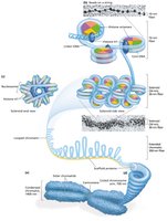
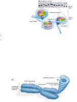
Least condensed chromatin appears as a 10-nm fiber (“beads-on-a-string”), with nucleosomes as beads and linker DNA as string.
Higher-Order Chromatin Organization and Chromosome Structure
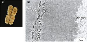
Chromatin forms 30-nm fibers during interphase and becomes maximally condensed in metaphase. Loops of 30-nm fibers attach to a chromosome scaffold, forming 300-nm fibers and giving chromosomes their shape.
Nucleosome Disassembly, Synthesis, and Reassembly During Replication
During DNA replication, old histones are retained as single molecules, dimers, or tetramers. New nucleosomes are assembled from both old and new histones.
Nucleosome Dynamics During Transcription
Transcription requires chromatin remodeling to convert chromatin from a closed (condensed) to an open (accessible) state. Remodeling complexes and transcription factors slide or eject nucleosomes, allowing RNA polymerase access. After transcription, nucleosomes are restored, silencing the gene.
Key Equations and Concepts
Chromosome Number: for diploid organisms, where is the number of unique chromosomes.
Aneuploidy: Trisomy (), Monosomy ().
Polyploidy: Triploid (), Tetraploid (), etc.
Example Table: Types of Chromosome Abnormalities
Type | Description | Example |
|---|---|---|
Aneuploidy | Abnormal number of chromosomes | Trisomy 21 (Down syndrome) |
Polyploidy | Extra sets of chromosomes | Triploid plants |
Deletion | Loss of chromosome segment | Cri du chat syndrome |
Duplication | Extra copy of chromosome segment | Charcot-Marie-Tooth disease |
Inversion | Reversed segment within chromosome | Paracentric inversion |
Translocation | Segment moved to another chromosome | Robertsonian translocation |
Additional info: Table entries and some examples are inferred for completeness and academic context.
