 Back
BackEukaryotic DNA Replication: Mechanisms, Enzymes, and Chromosome End Maintenance
Study Guide - Smart Notes
 Tailored notes based on your materials, expanded with key definitions, examples, and context.
Eukaryotic DNA Replication
Overview and Cell Cycle Context
DNA replication in eukaryotes is a highly regulated process that ensures the accurate duplication of the genome prior to cell division. It occurs during the S phase of the cell cycle, providing identical DNA copies for mitosis and meiosis.
Cell Cycle Phase: DNA replication occurs exclusively during the S (synthesis) phase.
Purpose: To produce two identical sets of chromosomes for daughter cells.
Semiconservative Mechanism: Each new DNA molecule consists of one parental and one newly synthesized strand.
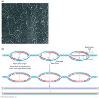
Multiple Origins and Replication Bubbles
Eukaryotic chromosomes are linear and contain multiple origins of replication, allowing for efficient and simultaneous DNA synthesis across large genomes. Replication proceeds bidirectionally from each origin, forming replication bubbles that eventually merge.
Replication Origin: Specific DNA sequences where replication begins.
Replication Bubble: The region of locally unwound DNA where replication occurs.
Bidirectional Synthesis: Two replication forks move in opposite directions from each origin.

Mechanism of DNA Replication
Stages of Replication
DNA replication can be divided into three main stages: initiation, elongation, and termination. Each stage involves specific enzymes and protein complexes.
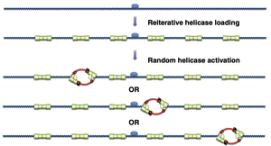
Initiation: Begins at multiple origins with the assembly of initiator proteins and loading of helicase.
Elongation: DNA synthesis proceeds in the 5′→3′ direction on both strands, with leading and lagging strand synthesis coordinated at the replication fork.
Termination: RNA primers are removed, DNA fragments are joined, and replication is completed.
Key Enzymes and Protein Complexes
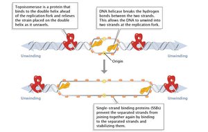
Multiple enzymes and protein complexes coordinate the unwinding of DNA, stabilization of single strands, and synthesis of new DNA.
Origin Recognition Complex (ORC): Binds to replication origins and recruits additional factors.
MCM Helicase: Unwinds the DNA double helix by breaking hydrogen bonds.
Topoisomerase: Relieves supercoiling and torsional strain ahead of the replication fork.
Single-Stranded Binding Proteins (SSBs): Stabilize unwound DNA and prevent reannealing.
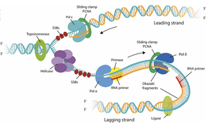
DNA Polymerases and Strand Synthesis
Three main DNA polymerases are involved in eukaryotic DNA replication, each with specialized roles:
DNA Polymerase α (alpha)–Primase: Synthesizes a short RNA primer and extends it with a short DNA segment.
DNA Polymerase ε (epsilon): Synthesizes the leading strand continuously.
DNA Polymerase δ (delta): Synthesizes the lagging strand discontinuously as Okazaki fragments.
Sliding Clamp (PCNA) and Clamp Loader (RFC): Ensure high processivity by tethering polymerases to DNA.
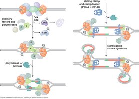
Leading and Lagging Strand Synthesis
DNA synthesis is continuous on the leading strand and discontinuous on the lagging strand, resulting in Okazaki fragments that are later joined.
Leading Strand: Synthesized continuously in the direction of fork movement by DNA polymerase ε (epsilon).
Lagging Strand: Synthesized discontinuously in short Okazaki fragments by DNA polymerase δ (delta).
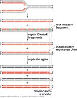
Primer Removal: RNase H removes RNA primers; DNA polymerase δ fills in gaps; DNA ligase seals nicks.

Comparison: Eukaryotic vs. Prokaryotic DNA Replication
While the core mechanism of DNA replication is conserved, several key differences exist between eukaryotes and prokaryotes:
Feature | Eukaryotes | Prokaryotes |
|---|---|---|
Chromosome Structure | Linear | Circular |
Number of Origins | Multiple per chromosome | Single per chromosome |
Polymerase Organization | Multiple specialized polymerases | Single main polymerase (Pol III) |
End-Replication Problem | Present (requires telomerase) | Absent (no ends to shorten) |
End-Replication Problem and Telomere Maintenance
The End-Replication Problem
Linear chromosomes present a unique challenge: after removal of the final RNA primer on the lagging strand, DNA polymerase cannot fill in the gap, leading to progressive chromosome shortening with each cell division.
Result: Unreplicated single-stranded region at the 3′ end of the template strand.
Consequence: Chromosome ends (telomeres) shorten with each replication cycle.
Telomeres and Telomerase
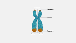
Telomeres are repetitive DNA sequences at chromosome ends that protect genetic material from degradation. Telomerase is an enzyme that extends telomeres, counteracting the end-replication problem.
Telomere Structure: Short, tandem repeats (e.g., TTAGGG in vertebrates) at chromosome termini.
Telomerase: A ribonucleoprotein enzyme with an RNA template and reverse transcriptase activity.
Function: Adds telomeric repeats to the 3′ end, allowing complete replication of chromosome ends.
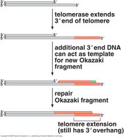
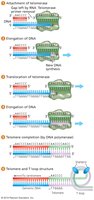
Mechanism of Telomerase Action
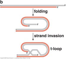
Telomerase binds to the 3′ overhang, uses its RNA template to extend the DNA, and allows primase and DNA polymerase δ to fill in the complementary strand. The telomere forms a protective T-loop structure stabilized by shelterin proteins.
Extension: Telomerase adds multiple telomeric repeats to the 3′ end.
Filling In: Primase and DNA polymerase δ synthesize the complementary strand.
T-Loop Formation: The extended telomere folds back, protecting chromosome ends.

Summary Table: Key Enzymes and Functions in Eukaryotic DNA Replication
Enzyme/Protein | Function |
|---|---|
ORC (Origin Recognition Complex) | Binds replication origins, recruits initiator proteins |
MCM Helicase | Unwinds DNA double helix |
Topoisomerase | Relieves supercoiling ahead of fork |
SSBs | Stabilize single-stranded DNA |
Primase | Synthesizes RNA primers |
DNA Polymerase α | Extends RNA primer with short DNA segment |
DNA Polymerase ε | Synthesizes leading strand |
DNA Polymerase δ | Synthesizes lagging strand |
RNase H | Removes RNA primers |
DNA Ligase | Seals nicks between Okazaki fragments |
PCNA (Sliding Clamp) | Increases polymerase processivity |
Telomerase | Extends telomeres to solve end-replication problem |
Key Equations and Concepts
Semiconservative Replication: Each daughter DNA molecule contains one parental and one new strand.
Direction of Synthesis: DNA is always synthesized in the 5′→3′ direction.
Telomere Repeat Sequence (Vertebrates):
Example: Application of DNA Replication Concepts
Example: In a rapidly dividing human cell, DNA replication must be completed efficiently and accurately. Multiple origins of replication ensure that the entire genome is duplicated within the limited time of S phase. Telomerase activity in stem cells and germ cells maintains telomere length, preventing loss of genetic information over successive divisions.
