 Back
BackCh 14 P1 Gene Mutation, DNA Repair, and Transposition: Mechanisms and Consequences
Study Guide - Smart Notes
 Tailored notes based on your materials, expanded with key definitions, examples, and context.
Ch 14 P1 Gene Mutation, DNA Repair, and Transposition
Introduction to Mutation
Mutations are permanent alterations in the nucleotide sequence of the genome. They can range from single base-pair changes to large chromosomal rearrangements. Mutations may occur in coding or noncoding regions, including regulatory sequences such as promoters, enhancers, and splicing signals.
Mutation: Any change in the DNA sequence, including substitutions, insertions, deletions, or chromosomal alterations.
Location: Mutations can affect gene function depending on their position within the genome.
Types of Mutations by Molecular Change
Mutations are classified based on the nature of the molecular change they introduce into the DNA sequence.
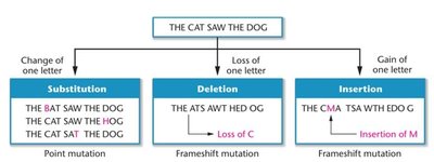
Point mutation (Base substitution): Change from one base pair to another.
Missense mutation: Alters a codon, resulting in a different amino acid.
Nonsense mutation: Converts a codon into a stop codon, leading to premature termination of translation.
Silent mutation: Alters a codon but does not change the encoded amino acid.
Neutral mutation: Occurs in noncoding regions and typically does not affect gene function.
Frameshift Mutations
Frameshift mutations result from the insertion or deletion of nucleotides, causing a shift in the reading frame during translation. This often leads to the production of nonfunctional proteins due to the introduction of premature stop codons.
Insertion/Deletion: Addition or loss of nucleotides not in multiples of three disrupts the triplet reading frame.
Effect: Can result in extensive missense or nonsense sequences downstream of the mutation.
Classification Based on Effect on Function
Mutations can be categorized by their impact on gene function and phenotype.
Loss-of-function mutation: Reduces or eliminates the function of the gene product.
Null mutation: Complete loss of gene function.
Recessive mutation: Typically results in loss of function and is masked in heterozygotes.
Dominant mutation: Produces a mutant phenotype in heterozygotes.
Dominant negative mutation: Mutant allele interferes with the function of the wild-type allele (often due to haploinsufficiency).
Gain-of-function mutation: Results in a gene product with enhanced, new, or negative functions; usually dominant.
Suppressor mutation: A second mutation that counteracts the effects of a previous mutation (can be intragenic or intergenic).
Classification Based on Location
Mutations are also classified by their location within the organism or genome.
Somatic mutations: Occur in non-germ cells; not heritable.
Germ-line mutations: Occur in gametes; heritable and passed to offspring.
Autosomal mutations: Affect genes on autosomes.
X-linked and Y-linked mutations: Affect genes on sex chromosomes.
Spontaneous and Induced Mutations
Mutations can arise spontaneously or be induced by external factors.
Spontaneous mutations: Occur naturally, often during DNA replication.
Induced mutations: Result from exposure to mutagens such as radiation or chemicals.
Mutation Rates
The mutation rate is the probability that a gene will mutate in a single generation or gamete. Mutation rates are generally low but vary among organisms and genes, reflecting differences in DNA proofreading and repair mechanisms.
Viral/Bacterial genes: Mutation rate ~1 in 100 million.
Human genome: Each newborn has ~60 new mutations compared to parents; number increases with paternal age.

Mechanisms of Spontaneous Mutation
Several molecular mechanisms contribute to spontaneous mutations.
Replication slippage: DNA polymerase slips during replication, especially in repeat sequences, leading to insertions or deletions. Associated with diseases like Fragile-X and Huntington disease.
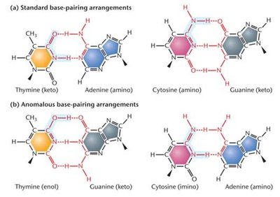
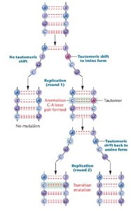
Tautomeric shifts: Temporary changes in base structure (tautomers) increase the likelihood of mispairing during replication, resulting in base substitutions.
Mutagens and Induced Mutations
Mutagens are agents that increase the frequency of mutations. They can be physical (e.g., radiation) or chemical (e.g., base analogs, alkylating agents).
Physical mutagens: UV light, X-rays, gamma rays, cosmic rays.
Chemical mutagens: Base analogs, alkylating agents, intercalating agents, adduct-forming agents.
Oxidative stress: Reactive oxygen species (ROS) cause base modifications, loss, or strand breaks.
Base Analogs
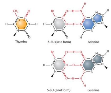
Base analogs are chemicals that resemble DNA bases and can be incorporated during replication, increasing mutation rates. For example, 5-bromouracil (5-BU) can pair with adenine or guanine, depending on its tautomeric form.
Alkylating, Intercalating, and Adduct-Forming Agents
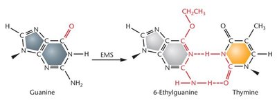
Alkylating agents: Add alkyl groups to bases, altering base-pairing and causing transition mutations (e.g., ethyl methane sulfonate).
Intercalating agents: Insert between base pairs, causing DNA distortion and frameshift mutations.
Adduct-forming agents: Covalently bind to DNA, altering its structure and interfering with replication and repair (e.g., acetaldehyde, heterocyclic amines).
Ultraviolet Light (UV) and Ionizing Radiation
UV light and ionizing radiation are potent physical mutagens.
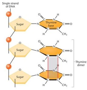
UV light: Causes formation of pyrimidine dimers (especially thymine dimers), distorting DNA and blocking replication/transcription.
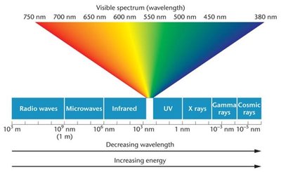
Ionizing radiation: Includes X-rays, gamma rays, and cosmic rays. Penetrates tissues, creates free radicals, and causes DNA breaks, deletions, and chromosomal rearrangements.
Genetic Diseases and Mutation Types
Genetic diseases can be polygenic (involving multiple genes) or monogenic (caused by a single gene mutation). Monogenic diseases often result from specific types of mutations, as summarized below.
Type of Mutation | Disorder | Molecular Change |
|---|---|---|
Missense | Achondroplasia | Glycine to arginine at position 380 of FGFR3 gene |
Nonsense | Marfan syndrome | Tyrosine to Stop codon at position 2113 of fibrillin-1 gene |
Insertion | Familial hypercholesterolemia | Various short insertions throughout the LDLR gene |
Deletion | Cystic fibrosis | Three-base-pair deletion of phenylalanine codon at position 508 of CFTR gene |
Trinucleotide repeat expansions | Huntington disease | >40 repeats of (CAG) sequence in coding region of Huntingtin gene |
Single-Gene Mutations and β-Thalassemia
β-Thalassemia is a monogenic, autosomal blood disorder caused by mutations in the β-globin gene (HBB). Over 400 mutations are known, affecting various gene regions and leading to a spectrum of disease severity.
Gene Region Affected | Number of Mutations Known | Description |
|---|---|---|
5′ upstream region | 22 | Single base-pair mutations between −101 and −25 upstream from transcription start site; e.g., T→A in TATA box at −30 reduces transcription. |
mRNA CAP site | 1 | A→C at +1 position decreases mRNA levels. |
5′ untranslated region | 3 | Mutations at +20, +22, +33 decrease transcription/translation, causing mild disease. |
ATG translation initiation codon | 7 | Mutations alter AUG, preventing translation and causing severe disease. |
Exons 1, 2, 3 | 36 | Missense, nonsense, and splice site mutations; severity varies. |
Introns 1 and 2 | 38 | Transitions/transversions reduce or abolish splicing, causing severe disease. |
Polyadenylation site | 6 | Changes in AATAAA reduce mRNA cleavage/polyadenylation, yielding unstable mRNAs (mild disease). |
Throughout/surrounding HBB | >100 | Insertions, deletions, duplications causing frameshifts, stop codons, and splicing defects. |
