 Back
BackGenes, Proteins, and Human Disease: The Molecular Basis of Heredity and Variation
Study Guide - Smart Notes
 Tailored notes based on your materials, expanded with key definitions, examples, and context.
Genes, Proteins, and Human Disease
Introduction to the Molecular Basis of Heredity
Understanding what genes do is central to genetics. Genes are responsible for encoding the information necessary to produce proteins, which in turn determine the phenotype of an organism. Mutations in genes can disrupt normal biological processes and lead to disease.
Historical Foundations: Genes and Metabolic Diseases
Archibald Garrod and Inborn Errors of Metabolism
In 1902, Archibald Garrod described alkaptonuria, a disease characterized by black urine and arthritis, caused by the accumulation of homogentisic acid. Garrod proposed that this was an 'inborn error of metabolism'—a genetic, not infectious, disease. He later showed the disease runs in families and suggested it was a recessive Mendelian trait, providing early evidence that genes control specific chemical reactions in the body.

Defining Genes: Genetic and Biochemical Perspectives
What is a Gene?
Genetic definition: A gene controls some aspect of an organism’s phenotype and segregates in defined ways between parents and offspring.
Biochemical definition: A gene is a segment of DNA that contains the information to express a protein (or sometimes RNA) that performs a function in the cell or body.
Proteins: Structure and Function
Proteins as Polymers of Amino Acids
Proteins are linear chains of amino acids, each with a unique sequence. The sequence of amino acids determines the protein’s structure and function.
Amino acids: There are 20 common amino acids, each with a distinct side chain (R group) that determines its chemical properties.
Peptide bond: Amino acids are joined by peptide bonds, forming polypeptide chains with an amino (N) terminus and a carboxyl (C) terminus.
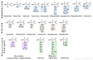
Hydrophilic and Hydrophobic Amino Acids
The chemical nature of the R group allows amino acids to be classified as hydrophilic (water-loving) or hydrophobic (water-fearing). Hydrophilic amino acids are typically found on the outside of proteins, while hydrophobic amino acids are buried inside.
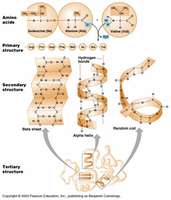
Levels of Protein Structure
Proteins fold into precise three-dimensional structures, which are critical for their function. There are four levels of protein structure:
Primary structure: The linear sequence of amino acids joined by covalent bonds.
Secondary structure: Local structures (e.g., alpha helices, beta sheets) formed by hydrogen bonding between nearby amino acids.
Tertiary structure: The overall 3D shape formed by long-range interactions, including hydrophobic/hydrophilic effects and ionic bonds.
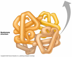
Quaternary structure: The assembly of multiple polypeptide chains into a functional protein complex (e.g., hemoglobin).
Genes, Proteins, and Enzymes
Enzymes as Biological Catalysts
Most genes encode proteins, and many of these proteins are enzymes. Enzymes catalyze specific chemical reactions in the cell, often working in pathways to synthesize or degrade molecules. The active site of an enzyme is where the chemical reaction occurs.
Biochemical Pathways and Disease
Enzymes function in metabolic pathways. If an enzyme is missing due to a gene mutation, its substrate may accumulate, leading to disease. For example, in alkaptonuria, a defective enzyme in the phenylalanine breakdown pathway leads to the accumulation of homogentisic acid.
Beadle and Tatum: The One Gene–One Enzyme Hypothesis
Neurospora crassa as a Model Organism
Beadle and Tatum used the bread mold Neurospora crassa to show that each gene controls a specific step in a biochemical pathway. They exposed spores to mutagens, creating auxotrophic mutants that could not synthesize specific vitamins or amino acids.
Prototroph: Can grow on minimal medium; synthesizes all required compounds.
Auxotroph: Cannot grow on minimal medium; requires supplementation with a specific compound.
By supplementing media with different nutrients, they identified which step in the pathway was blocked, leading to the 'one gene–one enzyme' hypothesis (later refined to 'one gene–one polypeptide').
Human Genetic Diseases: Enzyme Deficiencies and Protein Defects
Phenylketonuria (PKU)
PKU is caused by mutations in the gene encoding phenylalanine hydroxylase (PAH). Without PAH, phenylalanine accumulates and is converted to harmful products, leading to intellectual disability if untreated. Early detection and dietary management can prevent symptoms.
Tay-Sachs Disease
Tay-Sachs is due to a defect in the lysosomal enzyme HEXA, leading to the accumulation of a substrate in neurons. This causes neurodegeneration, paralysis, and early death. The disease is more common in certain populations due to genetic drift and founder effects.
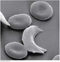
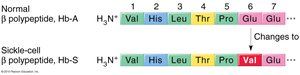
Sickle Cell Anemia: A Molecular Disease
Sickle cell anemia is caused by a mutation in the beta-globin gene, resulting in a single amino acid substitution (glutamic acid to valine at position 6, E6V). This change alters hemoglobin structure, causing red blood cells to adopt a sickle shape under low oxygen conditions. Sickled cells are fragile and can block capillaries, leading to pain, anemia, and organ damage.
Protein Electrophoresis and Genotype Identification
Protein gel electrophoresis can distinguish between normal and sickle cell hemoglobin based on differences in charge and shape, allowing for genotype identification.
Population Genetics: Sickle Cell and Malaria
The sickle cell allele persists at high frequency in populations where malaria is endemic because heterozygotes (carriers) have increased resistance to malaria. This is an example of balanced polymorphism due to heterozygote advantage.
Summary Table: Key Genetic Diseases and Their Molecular Basis
Disease | Gene/Protein Affected | Molecular Defect | Phenotype |
|---|---|---|---|
Alkaptonuria | Homogentisate 1,2-dioxygenase | Enzyme deficiency in phenylalanine breakdown | Black urine, arthritis |
Phenylketonuria (PKU) | Phenylalanine hydroxylase (PAH) | Enzyme deficiency, phenylalanine accumulation | Intellectual disability, musty odor |
Tay-Sachs Disease | HEXA (lysosomal enzyme) | Enzyme deficiency, substrate accumulation in neurons | Neurodegeneration, early death |
Sickle Cell Anemia | Beta-globin (hemoglobin) | Single amino acid substitution (E6V) | Sickled RBCs, anemia, pain |
Key Concepts and Equations
Central Dogma: Genetic information flows from DNA → RNA → Protein.
Peptide bond formation:
Genotype to phenotype: A single base change in DNA can lead to a single amino acid change in a protein, potentially altering its function and causing disease.
Conclusion
Genes encode proteins that determine the structure and function of cells. Mutations in genes can disrupt protein function, leading to metabolic and structural diseases. The study of these relationships forms the foundation of molecular genetics and has profound implications for medicine and evolutionary biology.
