 Back
BackGenes, Proteins, Enzymes, and Human Disease: Molecular Genetics Study Guide
Study Guide - Smart Notes
 Tailored notes based on your materials, expanded with key definitions, examples, and context.
Genes and Human Disease
Historical Discovery of Genetic Diseases
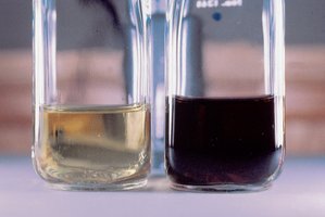
Mutations in genes can cause human diseases, as first described by Archibald Garrod in 1902 with the disease alkaptonuria. This disease is characterized by black urine and arthritis, resulting from the accumulation of homogentisic acid, which turns black upon exposure to air. Garrod termed this an 'inborn error of metabolism,' indicating a genetic rather than infectious origin.
Key Point: Alkaptonuria is inherited and not caused by germs.
Key Point: Garrod suggested it was a recessive Mendelian trait.
Key Point: Genes are required for specific chemical reactions in the body.
Example: Alkaptonuria results from a defective enzyme in the phenylalanine breakdown pathway.
What is a Gene?
Genetic and Biochemical Definitions
Genes have been defined both genetically and biochemically. Genetically, a gene controls some aspect of an organism’s phenotype and resides on chromosomes, segregating in defined ways. Biochemically, a gene is a segment of DNA containing the information to express a protein that performs a function in the cell or body.
Key Point: Genes determine form, function, and behavior.
Key Point: Genes encode proteins, which are the functional molecules in cells.
Proteins: Structure and Function
Amino Acids and Protein Structure
Proteins are polymers made of amino acids, each with a unique sequence. There are 20 common amino acids, grouped by their R side chains into categories: nonpolar, polar, acidic, and basic. The sequence and chemical properties of amino acids determine how a protein folds and functions.
Key Point: Proteins are linear chains of covalently joined amino acids.
Key Point: The peptide bond joins the carboxyl group of one amino acid to the amino group of another.
Key Point: Polypeptide chains have an amino terminus (start) and a carboxyl terminus (end).
Example: Hemoglobin protein sequence.


Grouping of Amino Acids
The chemical nature of the R group is used to group amino acids into categories. Charged and polar amino acids are hydrophilic and found on the outside of proteins, while nonpolar amino acids are hydrophobic and found on the inside.
Key Point: Hydrophilic amino acids are exposed to water; hydrophobic amino acids are sheltered inside.
Levels of Protein Structure
Proteins fold into precise three-dimensional structures based on their amino acid sequence. The structure determines the function performed in the cell. There are four levels of protein structure:
Primary structure: Linear sequence of amino acids joined by covalent bonds.
Secondary structure: Local structures formed by hydrogen bonds (alpha helix, beta sheet).
Tertiary structure: Final 3D shape formed by long-range interactions (hydrogen bonds, ionic bonds, hydrophobic/hydrophilic effects).
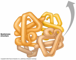
Quaternary structure: Multiple polypeptides working together (e.g., hemoglobin has 2 alpha and 2 beta subunits).

Genes, Proteins, and Enzymes
Enzymes as Proteins
Most genes contain instructions to make proteins, many of which are enzymes. Enzymes are proteins that catalyze specific chemical reactions. The active site of an enzyme is where the reaction occurs, and enzymes work together in pathways to synthesize or break down substances.
Key Point: Enzymes are proteins that fold to perform specific chemical reactions.
Key Point: Enzymes can break down or synthesize molecules.
Key Point: Enzymes work in pathways; missing enzymes cause substrate accumulation.
Biochemical Pathways and Genetic Diseases
Alkaptonuria and Enzyme Deficiency
Alkaptonuria is caused by a defective enzyme in the phenylalanine breakdown pathway, leading to the accumulation of homogentisic acid. In biochemical pathways, if an enzyme is missing, its substrate accumulates, resulting in disease.
Key Point: Enzyme defects in pathways cause metabolic diseases.
Example: Alkaptonuria results from lack of enzyme to break down homogentisic acid.
Beadle and Tatum: One Gene-One Enzyme Hypothesis
Model Organisms and Genetic Screens
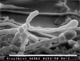
Beadle and Tatum used the bread mold Neurospora crassa to show that one gene controls one enzyme at a specific step in a biochemical pathway. They exposed spores to mutagens to create auxotrophic mutants, which could not synthesize specific vitamins or amino acids.
Key Point: Prototrophs can grow on minimal media; auxotrophs require supplements.
Key Point: Genetic screens identify mutants with defects in biochemical pathways.
Example: Mutants unable to synthesize methionine or arginine.
Biochemical Pathway Analysis
By supplementing media with specific compounds, Beadle and Tatum determined which step in a pathway was defective. Mutants could grow with arginine or citrulline but not ornithine, indicating a defect in the enzyme converting ornithine to citrulline.
Key Point: Mutations in single genes affect specific steps in pathways.
Key Point: Led to the 'one gene-one enzyme' hypothesis, later revised to 'one gene-one polypeptide.'
The Central Dogma of Molecular Genetics
Genotype to Phenotype
The central dogma describes the flow of genetic information: DNA is transcribed to RNA, which is translated to protein. Proteins determine cellular form and function, linking genotype to phenotype.
Key Point: DNA → RNA → Protein → Phenotype
Genetic Diseases: Enzyme and Protein Defects
Phenylketonuria (PKU)
PKU is caused by mutations in the gene encoding phenylalanine hydroxylase (PAH). Without PAH, phenylalanine accumulates and is converted to harmful products, leading to severe mental retardation. Early detection and dietary management prevent symptoms.
Key Point: PKU is a monogenic disease caused by enzyme deficiency.
Key Point: More than 400 recessive mutations in PAH identified.
Example: Newborn screening for PKU.
Tay-Sachs Disease
Tay-Sachs is caused by a defect in the lysosomal enzyme HEXA, leading to accumulation of its substrate and neuronal degeneration. It is common among Jewish people of European ancestry and has no cure, but genetic testing has reduced prevalence.
Key Point: Tay-Sachs is caused by enzyme deficiency in lysosomes.
Key Point: Symptoms include paralysis, blindness, and early death.
Sickle Cell Anemia: A Molecular Disease
Sickle cell anemia is caused by mutations in the gene for beta hemoglobin. Hemoglobin is composed of two alpha and two beta chains. A single amino acid change (E6V: glutamic acid to valine) alters the protein’s structure, causing red blood cells to adopt a sickle shape, leading to fragility and blockage of capillaries.
Key Point: Sickle cell anemia is due to a single amino acid change in hemoglobin.
Key Point: The disease is common in populations exposed to malaria.
Example: Protein gel electrophoresis distinguishes normal and mutant hemoglobin.

Protein Gel Electrophoresis
Protein gel electrophoresis separates proteins based on size, charge, and shape. Linus Pauling used this technique to show that sickle cell hemoglobin behaves differently from normal hemoglobin, identifying sickle cell anemia as a molecular disease.
Key Point: Gel electrophoresis can distinguish genotypes based on protein migration.
Sickle Cell Mutation and Malaria Resistance
People heterozygous for the sickle cell mutation (carriers) have increased resistance to malaria, as the altered red blood cells disrupt the life cycle of Plasmodium falciparum. This explains the high frequency of the sickle cell allele in populations where malaria is prevalent.
Key Point: Heterozygote advantage maintains sickle cell allele in malarial regions.
Key Point: Sickle cell disease is most common in populations with historical malaria exposure.
Summary Table: Amino Acid Properties
The 20 amino acids are grouped by their chemical properties, which influence protein folding and function.
Group | Amino Acids | Properties |
|---|---|---|
Nonpolar | Glycine, Alanine, Valine, Leucine, Isoleucine, Methionine, Tryptophan, Phenylalanine, Proline | Hydrophobic, found inside proteins |
Polar | Serine, Threonine, Cysteine, Tyrosine, Asparagine, Glutamine | Hydrophilic, found outside proteins |
Acidic | Aspartic acid, Glutamic acid | Negatively charged |
Basic | Lysine, Arginine, Histidine | Positively charged |
Summary Table: Sickle Cell Mutation
Position | Normal Hb-A | Sickle Cell Hb-S |
|---|---|---|
6 | Glu (Glutamic acid) | Val (Valine) |
Key Equations
Peptide Bond Formation:
Central Dogma:
Mutation Effect:
Additional info: Expanded explanations and context were added to clarify the molecular basis of heredity, gene function, protein structure, and the genetic basis of human diseases, as well as the historical experiments that established these principles.
