 Back
BackGenes, Proteins, Enzymes, and Human Disease: Molecular Genetics Study Guide
Study Guide - Smart Notes
 Tailored notes based on your materials, expanded with key definitions, examples, and context.
Genes and Human Disease
Historical Discovery of Genetic Diseases
Mutations in genes can cause human diseases, as first described by Archibald Garrod in 1902 with the disease alkaptonuria. This disease is characterized by black urine and arthritis, resulting from the accumulation of homogentisic acid (alkapton), which turns black upon exposure to air. Garrod termed this an 'inborn error of metabolism,' indicating a genetic rather than infectious origin.
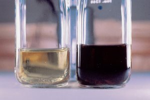
Key Point: Alkaptonuria is inherited and not caused by germs.
Key Point: Garrod suggested it was a recessive Mendelian trait.
Example: The disease runs in families, supporting genetic inheritance.
What is a Gene?
Genetic and Biochemical Definitions
Genes have been defined both genetically and biochemically. Genetically, a gene controls some aspect of an organism’s phenotype and resides on chromosomes, segregating in defined ways. Biochemically, a gene is a segment of DNA containing the information to express a protein that performs a function in the cell or body.
Key Point: Genes are the fundamental units of heredity and function.
Key Point: Most genes encode proteins, which carry out cellular functions.
Proteins: Structure and Function
Amino Acids and Protein Structure
Proteins are polymers made of amino acids, each with a unique sequence. There are 20 amino acids commonly found in proteins, grouped by their R side chains into categories: nonpolar, polar, acidic, and basic. The sequence and chemical properties of amino acids determine protein folding and function.
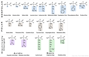
Key Point: Amino acids are joined by peptide bonds, forming polypeptide chains.
Key Point: Polypeptide chains have an amino terminus (start) and a carboxyl terminus (end).
Example: The hemoglobin protein begins with a specific amino acid sequence.
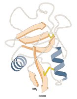
Levels of Protein Structure
Proteins fold into precise three-dimensional structures, which determine their function. There are four levels of protein structure:
Primary Structure: Linear sequence of amino acids joined by covalent bonds.
Secondary Structure: Local structures formed by hydrogen bonds (e.g., alpha helix, beta sheet).
Tertiary Structure: Final 3D shape formed by long-range interactions and hydrophobic/hydrophilic properties.
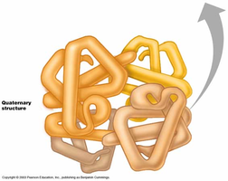
Quaternary Structure: Multiple polypeptides working together (e.g., hemoglobin has 2 alpha and 2 beta subunits).
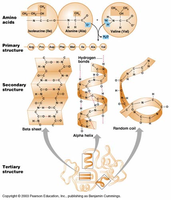
Additional info: The forces holding proteins in their conformation are weaker than covalent bonds, such as hydrogen bonds and ionic interactions.
Genes, Proteins, and Enzymes
Enzymes as Proteins
Most genes contain instructions to make proteins, and many proteins are enzymes. Enzymes are proteins that catalyze specific chemical reactions, folding to create an active site where the reaction occurs.
Key Point: Enzymes perform chemical reactions essential for life.
Key Point: Some enzymes break down molecules, others synthesize them.
Mutations and Biochemical Pathways
Alkaptonuria and Enzyme Deficiency
Alkaptonuria is caused by a defective enzyme in the pathway for breaking down phenylalanine, leading to the accumulation of homogentisic acid. Enzymes work together in pathways, and a missing enzyme causes substrate accumulation.
Key Point: Mutations in genes can disrupt biochemical pathways.
Example: Alkaptonuria results from a block in the phenylalanine breakdown pathway.
Beadle and Tatum: One Gene-One Enzyme Hypothesis
Beadle and Tatum used Neurospora crassa to show that one gene controls one enzyme at a specific step in a biochemical pathway. They exposed spores to mutagens, isolated auxotrophic mutants, and determined which compounds mutants could not synthesize.
Key Point: Auxotrophs cannot grow on minimal media and require supplementation.
Key Point: Mutations in single genes affect specific steps in pathways.
Example: Mutants unable to synthesize methionine grow only when methionine is added.
Additional info: The hypothesis evolved to 'one gene-one polypeptide' as not all genes encode enzymes.
Genes, Enzymes, and Human Disease
Phenylketonuria (PKU)
PKU is caused by mutations in the gene encoding phenylalanine hydroxylase (PAH). Without PAH, phenylalanine accumulates and is converted to harmful products, leading to severe mental retardation. Early detection and dietary management prevent symptoms.
Key Point: PKU is a monogenic disease with over 400 known mutations.
Example: Newborns are screened for PKU and placed on a low-phenylalanine diet if affected.
Tay-Sachs Disease
Tay-Sachs is caused by a defect in the lysosomal enzyme HEXA, leading to substrate accumulation and neuronal degeneration. It is common among Jewish people of European ancestry and has no cure, but genetic testing has reduced prevalence.
Key Point: Tay-Sachs results in severe neurological symptoms and early death.
Example: Genetic screening helps prevent the disease in at-risk populations.
Sickle Cell Anemia: A Molecular Disease
Sickle cell anemia is caused by mutations in the gene for beta hemoglobin. Hemoglobin is composed of two alpha and two beta chains. A single amino acid change (E6V: glutamic acid to valine) alters hemoglobin's structure, causing red blood cells to adopt a sickle shape under low oxygen.
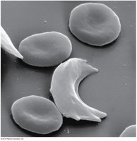
Key Point: Sickle cells are fragile and can block capillaries, leading to reduced oxygen transport.
Key Point: Sickle cell anemia is common in populations with malaria due to heterozygote advantage.
Example: Linus Pauling described sickle cell anemia as a 'molecular disease' based on protein gel electrophoresis.
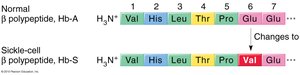
Additional info: A single base pair change in DNA can cause a single amino acid change in a protein, drastically altering its function.
Protein Structure and Disease
Protein Folding and Function
The three-dimensional structure of a protein is determined by its amino acid sequence and chemical properties. Mutations that alter this sequence can disrupt folding and function, leading to disease.
Key Point: Protein structure is essential for function; even a single amino acid change can have dramatic effects.
Example: Sickle cell anemia is caused by a single amino acid substitution.
Summary Table: Amino Acid Properties
The chemical nature of the R group is used to group amino acids into categories. This classification is important for understanding protein folding and function.
Category | Amino Acids | Properties |
|---|---|---|
Nonpolar | Glycine, Alanine, Valine, Leucine, Isoleucine, Methionine, Tryptophan, Phenylalanine, Proline | Hydrophobic, found inside proteins |
Polar | Serine, Threonine, Cysteine, Tyrosine, Asparagine, Glutamine | Hydrophilic, found on protein surfaces |
Acidic | Aspartic acid, Glutamic acid | Negatively charged |
Basic | Lysine, Arginine, Histidine | Positively charged |
Equations and Genetic Concepts
Key genetic equations and concepts relevant to protein structure and disease:
Peptide Bond Formation:
Central Dogma:
Mutation Impact:
Conclusion
Mutations in genes can cause human diseases by altering the structure and function of proteins, especially enzymes. Understanding the molecular basis of these diseases, such as alkaptonuria, PKU, Tay-Sachs, and sickle cell anemia, provides insight into the relationship between genotype and phenotype, and the importance of protein structure in cellular function.
