 Back
Genetic Code and Transcription: Structure, Function, and Mechanisms
Study Guide - Smart Notes
 Tailored notes based on your materials, expanded with key definitions, examples, and context.
Genetic Code and Transcription
The Central Dogma of Molecular Biology
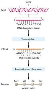
The central dogma describes the flow of genetic information within a biological system: DNA is transcribed into mRNA, which is then translated into protein. This process is fundamental to molecular genetics and cellular biology.
DNA contains the genetic instructions for all cellular functions.
Transcription is the process by which DNA is copied into messenger RNA (mRNA).
Translation is the process by which mRNA is decoded to synthesize proteins.
Proteins are the functional molecules responsible for cellular structure and activity.
Characteristics of the Genetic Code
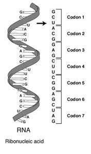
The genetic code is a set of rules by which information encoded in DNA is translated into proteins. It is linear, using nucleotide 'letters' (A, C, G, T/U) to form codons, which specify amino acids.
Codon: A sequence of three nucleotides in mRNA that codes for a specific amino acid.
Triplet code: Each amino acid is specified by a set of three nucleotides.
There are four nucleotides in RNA (A, C, G, U), allowing for 64 possible codons ().
Only 20 amino acids are encoded, so the code is degenerate (multiple codons per amino acid).
Discovery of mRNA and the Triplet Code
Early studies hypothesized a direct DNA-to-protein relationship, but evidence showed an unstable RNA intermediate. Jacob and Monod proposed the existence of messenger RNA (mRNA). Sydney Brenner postulated the triplet code, and Francis Crick's frameshift studies confirmed its necessity.
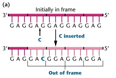
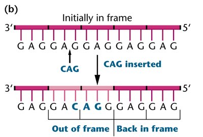
Frameshift mutations: Insertions or deletions of nucleotides that shift the reading frame, altering the resulting protein.
Only insertions/deletions in multiples of three nucleotides restore the reading frame.
Codon Assignment and Experimental Approaches
To determine which codons specify which amino acids, researchers used cell-free systems and synthetic mRNAs. Homopolymers (single nucleotide repeats) and mixed heteropolymers (combinations of nucleotides) were used to analyze codon-amino acid relationships.
Homopolymers: Strings of a single nucleotide (e.g., UUU codes for phenylalanine).
Mixed heteropolymers: Combinations of two or more nucleotides at known ratios to deduce codon assignments.
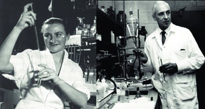
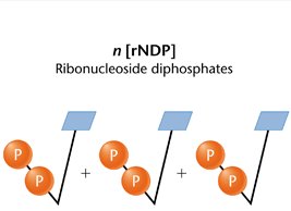
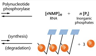
Polynucleotide phosphorylase: Enzyme used to synthesize RNA without a DNA template.
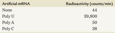
Homopolymer Experiment Results
Artificial mRNA | Radioactivity (counts/min) |
|---|---|
None | 44 |
Poly U | 39,800 |
Poly A | 50 |
Poly C | 38 |
Mixed Heteropolymer Analysis
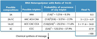
Possible compositions | Possible triplets | Probability of occurrence of any triplet | Final % |
|---|---|---|---|
3A | AAA | 0.5% | 0.5 |
2A:1C | AAC ACA CAA | 2.3% | 6.9 |
1A:2C | ACC CAC CCA | 11.6% | 34.8 |
3C | CCC | 57.9% | 57.9 |
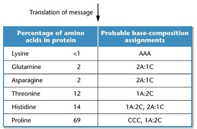
Translation of Message: Amino Acid Assignments
Percentage of amino acids in protein | Probable base-composition assignments |
|---|---|
Lysine | <1 | AAA |
Glutamine | 2 | 2A:1C |
Asparagine | 2 | 2A:1C |
Threonine | 12 | 1A:2C |
Histidine | 14 | 1A:2C, 2A:1C |
Proline | 69 | CCC, 1A:2C |
Degeneracy and Unambiguity of the Genetic Code
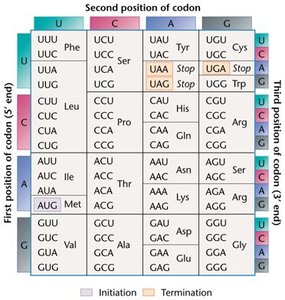
The genetic code is degenerate, meaning most amino acids are specified by more than one codon. However, each codon is unambiguous, coding for only one amino acid.
Leucine has six codons; tryptophan has only one.
18 of 20 amino acids have multiple codons.
Triplet Binding Assay
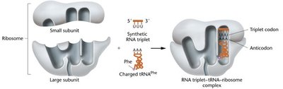
This assay uses synthetic RNA triplets to bind ribosomes and radiolabeled tRNAs, allowing identification of codon-amino acid pairs.
50 codons were determined using this method.
Repeating Copolymer Analysis
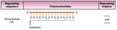
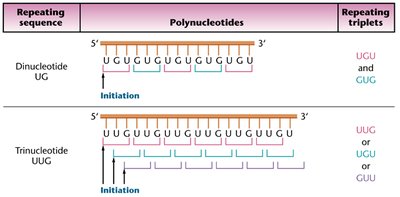
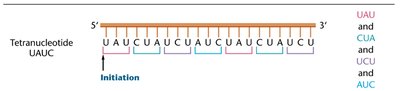
Longer synthetic RNA molecules with repeating di-, tri-, or tetra-nucleotide sequences were used to identify codons and confirm previous assignments. This method also revealed stop codons.
Frames such as UCU, CUC, UUA, UAC, ACU, and CUU were analyzed for their amino acid products.
Stop codons were identified when specific codons failed to incorporate proteins.
Wobble Hypothesis
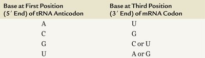
Francis Crick postulated that the first two nucleotide positions in a codon are more important than the third, allowing for flexible pairing (wobble) between tRNA anticodon and mRNA codon. This reduces the number of tRNAs needed.
Only 30 different tRNAs are needed in prokaryotes; eukaryotes have 41-55.
Start and Stop Codons
Specific codons signal the initiation and termination of translation. AUG is the start codon, coding for methionine. UAA, UAG, and UGA are stop codons, not recognized by tRNA, and signal the end of translation.
Start codon: AUG (sometimes GUG or UUG)
Stop codons: UAA, UAG, UGA
Nonsense mutation: Internal mutation causing a stop codon
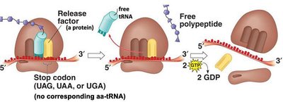
Universality and Colinearity of the Genetic Code
The genetic code is nearly universal across viruses, prokaryotes, and eukaryotes. mRNA and amino acid sequences are colinear, meaning they are read in the same direction without overlaps or breaks.
Each nucleotide belongs to only one codon.
No breaks in translation between codons, assuming no stop codon.
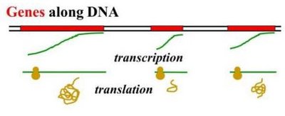
Transcription: DNA to mRNA
Components and Mechanism of Bacterial Transcription
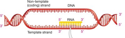
Transcription in bacteria involves the transcription holoenzyme, composed of sigma factor and RNA polymerase. The sigma factor recognizes the promoter sequence, and RNA polymerase links nucleotides to form mRNA.
Promoter region: Start of transcription (+1 site), includes -10 box (TATAAT) and -35 box (TTGACA).
Initiation: Sigma binds to promoter, RNA polymerase opens DNA helix.
Elongation: RNA polymerase synthesizes mRNA.
Termination: Hairpin loop forms in RNA, causing RNA and RNA polymerase to separate.
Comparison of Bacterial and Eukaryotic Transcription
Bacterial and eukaryotic transcription differ in the number of RNA polymerases, promoter structure, and associated proteins.
Bacteria: One RNA polymerase, -10 and -35 promoter boxes, sigma factor.
Eukaryotes: Three RNA polymerases (for different RNA types), variable promoter structure (often -30 TATA box), multiple basal transcription factors.
mRNA Processing in Eukaryotes
Structure of Eukaryotic Genes
Eukaryotic genes contain exons (coding regions) and introns (non-coding regions). Introns are removed during RNA splicing, and exons are joined to form mature mRNA.
Exons: Portions that get transcribed and translated.
Introns: Portions that are transcribed but not translated.
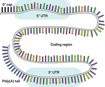
RNA Splicing and Modifications
RNA splicing is performed by the spliceosome, a complex of small nuclear RNA (snRNA) and proteins (snRNPs). Additional modifications include the addition of a 5' cap and a polyA tail.
5' cap: Enables ribosome binding and protects mRNA from degradation.
PolyA tail: Added to the 3' end, stabilizes mRNA.
Summary Table: Key Features of the Genetic Code
Feature | Description |
|---|---|
Triplet code | Three nucleotides per codon |
Degeneracy | Multiple codons per amino acid |
Unambiguity | Each codon specifies only one amino acid |
Universality | Nearly universal across organisms |
Colinearity | mRNA and protein sequences are aligned |
Start/Stop codons | AUG (start), UAA/UAG/UGA (stop) |
Key Equations
Number of possible codons:
Probability of a triplet in mixed heteropolymer: (for AAA in 1A:5C ratio)
Example: Codon Assignment
UUU codes for phenylalanine
AUG codes for methionine (start codon)
UAA, UAG, UGA are stop codons
Additional info:
Some images and tables were inferred to clarify experimental approaches and codon assignments.
Key historical figures and experiments were included for academic context.
