 Back
Genetic Material: Structure and Organization
Study Guide - Smart Notes
 Tailored notes based on your materials, expanded with key definitions, examples, and context.
Genetic Material: Structure and Organization
Introduction to Genetic Material
The genetic material is the foundation of heredity and biological information in all living organisms. It must be capable of replication, information storage, expression, and variation by mutation. These properties ensure faithful transmission of genetic information and enable diversity and evolution.
Replication: The genetic material must be copied accurately during cell division (mitosis and meiosis).
Information Storage: Genes serve as repositories of genetic instructions.
Expression: Genetic information must be expressed to produce functional molecules.
Variation: Mutations introduce genetic diversity, essential for evolution.
Historical Discovery of DNA as Genetic Material
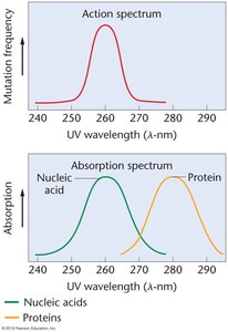
Early chemical studies identified DNA in chromosomes, but its role as genetic material was debated. Proteins, with their greater diversity, were initially considered more likely candidates. Key experiments demonstrated that DNA, not protein, is the substance of genes.
F. Meischer (1869): Extracted "nuclein" (DNA) from nuclei of human white blood cells.
Feulgen Reaction (1923): DNA localized almost exclusively within chromosomes.
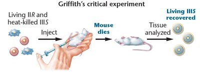
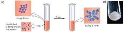
Griffith's Transformation Experiment (1928): Showed that genetic material from heat-killed virulent bacteria could transform nonvirulent bacteria.
Avery, MacLeod, and McCarty (1944): Identified DNA as the "transforming principle" responsible for genetic transformation.
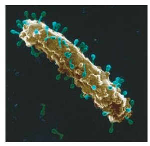
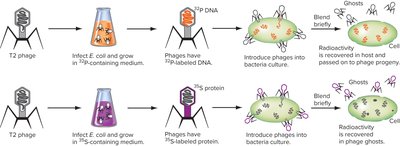
Hershey-Chase Experiment: Used bacteriophages to confirm that DNA, not protein, carries genetic information.
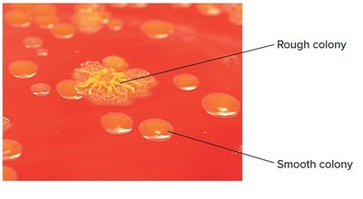
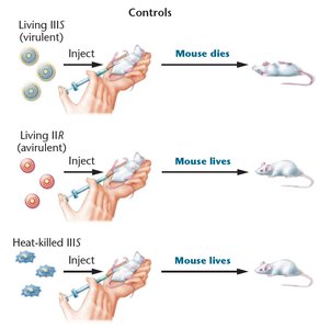
Serotype | Colony Morphology | Capsule | Virulence |
|---|---|---|---|
IIR | Rough | Absent | Avirulent |
IIIS | Smooth | Present | Virulent |
Chemistry of Genetic Material: Nucleosides and Nucleotides
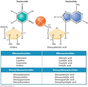
DNA and RNA are polymers of nucleotides, each consisting of a nitrogenous base, a pentose sugar, and a phosphate group. Nucleosides lack the phosphate group. The sequence of nucleotides encodes genetic information.
Nucleoside: Nitrogenous base + pentose sugar
Nucleotide: Nitrogenous base + pentose sugar + phosphate group
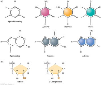
Types of bases: Purines (adenine, guanine) and pyrimidines (cytosine, thymine, uracil)
Polynucleotide Structure and Phosphodiester Bonds
Nucleotides are linked by phosphodiester bonds between the phosphate group at the C-5' position and the OH group at the C-3' position. This creates a backbone with distinct 5' and 3' ends, essential for directionality in DNA and RNA.
Phosphodiester bond: Connects nucleotides in a strand.
Oligonucleotides: Short chains (up to 30 nucleotides).
Polynucleotides: Long chains (>30 nucleotides).
Chargaff's Law and Base Composition
Chargaff's law states that in DNA, the amount of adenine (A) is approximately equal to thymine (T), and the amount of guanine (G) is approximately equal to cytosine (C). This pattern is essential for complementary base pairing and refuted earlier hypotheses of equal base distribution.
Base pairing: A pairs with T, G pairs with C.
Implication: Supports the double helix model and accurate replication.
DNA Structure: The Double Helix
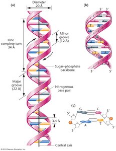
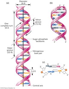
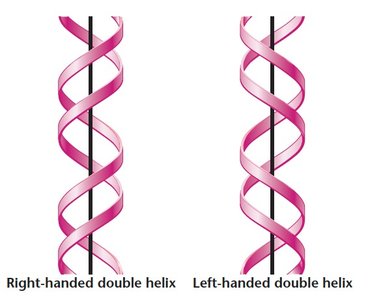
The Watson and Crick model (1953) describes DNA as a right-handed double helix composed of two antiparallel polynucleotide chains. The strands are held together by hydrogen bonds between complementary bases, with major and minor grooves along the helix.
Double helix: Two antiparallel strands, right-handed.
Base pairs: A-T and G-C, stacked 3.4 Å apart.
Helix dimensions: Diameter 20 Å, one turn 34 Å (10 base pairs).
Grooves: Major and minor grooves alternate along the helix.
Alternative Forms of DNA
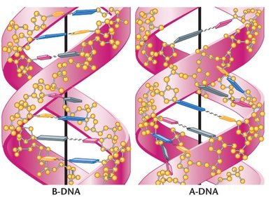
DNA can adopt several conformations depending on environmental conditions. The most common form in cells is B-DNA, but A-DNA, Z-DNA, and others exist under specific conditions.
B-DNA: Standard form under physiological conditions.
A-DNA: More compact, forms under high-salt or dehydration.
Z-DNA: Left-handed helix, forms under certain conditions.
C-, D-, E-, P-DNA: Less common, right-handed, some lack guanine.
DNA and Protein: Roles in Genetic Information
DNA stores genetic information, while proteins interpret, copy, regulate, and repair DNA. The sequence of bases in DNA encodes biological instructions, and complementary base pairing ensures accurate replication.
DNA: Stable storage of instructions.
Proteins: Read, copy, regulate, and repair DNA.
Cell Structure and DNA Organization
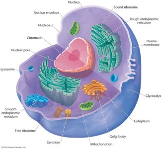
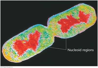
In eukaryotes, DNA is housed in the nucleus, organized into chromatin. The nucleolus is the site of rRNA synthesis. In prokaryotes, DNA is found in the nucleoid, which is not membrane-bound.
Nucleus: Membrane-bound, contains DNA.
Nucleolus: Site of rRNA synthesis.
Nucleoid: Prokaryotic DNA region, not membrane-bound.
Organization of DNA into Chromatin
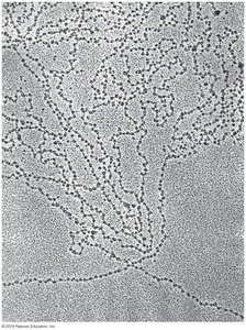
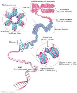
Chromatin is the complex of DNA and protein found in eukaryotic nuclei. During interphase, chromatin is dispersed; during cell division, it condenses into visible chromosomes. Chromatin consists of repeating units called nucleosomes, each containing DNA wrapped around histone proteins.
Chromatin: DNA-protein complex, dispersed in interphase, condensed in mitosis.
Nucleosomes: DNA wrapped around eight histone proteins (~200 bp per nucleosome).
Histones: Positively charged proteins (H1, H2A, H2B, H3, H4) facilitate DNA packaging.
Types of Chromatin
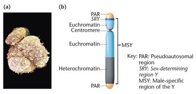
Chromatin can be classified as euchromatin or heterochromatin. Euchromatin contains functional genes and is actively transcribed, while heterochromatin is genetically inactive and replicates later in S phase.
Euchromatin: Contains functional genes, actively transcribed.
Heterochromatin: Genetically inactive, contains repressed genes, includes telomeres and centromeres.
Homologous Chromosomes
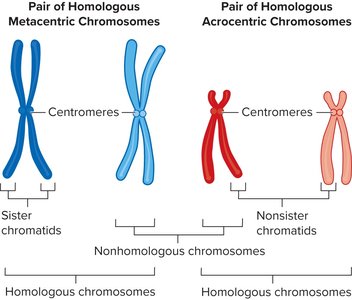
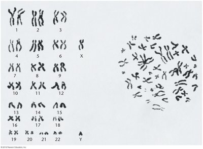
In diploid organisms, chromosomes exist in homologous pairs. Homologous chromosomes have identical gene loci but may carry different alleles. The centromere's location determines chromosome appearance and classification.
Homologous chromosomes: Similar but not identical, carry genes for same traits.
Alleles: Different versions of the same gene.
Centromere: Constricted region, determines chromosome type.
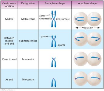
Centromere location | Designation | Metaphase shape | Anaphase shape |
|---|---|---|---|
Middle | Metacentric | Sister chromatids, centromere in middle | Migration |
Between middle and end | Submetacentric | p arm, q arm | Migration |
Close to end | Acrocentric | Centromere near end | Migration |
At end | Telocentric | Centromere at end | Migration |
Chromosome Number and Variation
Haploid chromosome numbers vary widely among organisms and do not correlate with organism size or complexity. Odd diploid numbers often result from hybridization or unusual chromosome systems.
Haploid number (n): Number of chromosomes in gametes.
Diploid number (2n): Number of chromosomes in somatic cells.
Hybridization: Can result in odd diploid numbers (e.g., mule).
Special Chromosomes
Some chromosomes exhibit unique structures and functions. Polytene chromosomes are paired homologs found in certain tissues and display puff regions of high gene activity. Lampbrush chromosomes are large with extensive DNA looping, found in oocytes and some insect spermatocytes.
Polytene chromosomes: Paired homologs, puff regions indicate active transcription.
Lampbrush chromosomes: Large, looped, sites of gene activity.
