 Back
BackGenetic Mechanisms: Double-Strand Break Repair, Transposable Elements, and Regulation of Gene Expression
Study Guide - Smart Notes
 Tailored notes based on your materials, expanded with key definitions, examples, and context.
Double-Strand Break Repair
Overview of Double-Strand Break Repair
Double-strand breaks (DSBs) in DNA are among the most severe forms of genetic damage, potentially leading to chromosomal rearrangements, cancer, or cell death. Cells have evolved two primary pathways to repair DSBs: homologous recombination repair and nonhomologous end joining.
Homologous recombination repair: Utilizes a homologous sequence as a template for accurate repair.
Nonhomologous end joining: Directly ligates the broken DNA ends, often resulting in mutations.
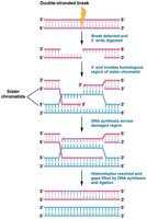
Homologous Recombination Repair
Homologous recombination repair is a high-fidelity mechanism that uses the sister chromatid as a template to restore the original DNA sequence. This process is most active during late S or early G2 phase when sister chromatids are available.
Recognition and resection: The break is recognized, and the 5' ends are digested, leaving 3' overhangs.
Strand invasion: The 3' overhang invades the homologous region of the sister chromatid.
DNA synthesis: DNA polymerase synthesizes new DNA using the sister chromatid as a template.
Resolution: The newly synthesized DNA is ligated, restoring the integrity of the chromosome.
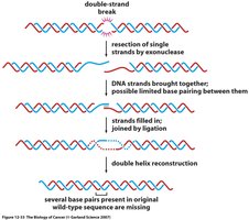
Nonhomologous End Joining (NHEJ)
Nonhomologous end joining is an error-prone repair mechanism that is active in G1 phase, prior to DNA replication. It involves a complex of proteins, including kinases and BRCA1, which bind to the free DNA ends and ligate them together.
Resection: Single strands are resected by exonucleases.
End joining: DNA ends are brought together, and limited base pairing may occur.
Ligation: Strands are filled in and joined by ligation, reconstructing the double helix.
Mutation risk: Several base pairs may be lost, leading to mutations.
Transposable Elements
Introduction to Transposable Elements
Transposable elements (TEs), also known as "jumping genes," are DNA sequences that can move within and between chromosomes. They are found in all organisms and can disrupt gene function or regulation.
Insertion sequences (IS elements): Simple transposons that cause mutations when inserted into genes.
Bacterial transposons: Larger elements that can spread drug resistance among bacteria.
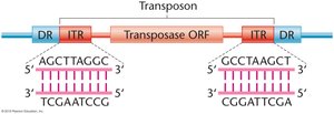
Structure of Transposons
Transposons typically contain inverted terminal repeats (ITRs) and direct repeats (DRs) flanking a transposase open reading frame (ORF).
ITRs: Inverted sequences at the ends of the transposon.
DRs: Direct repeats generated during insertion.
Transposase: Enzyme required for transposition.
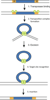
Mechanism of Transposition
Transposition involves several steps: binding of transposase, formation of a transposition complex, excision, target site recognition, and insertion.
Transposase binding: Recognizes and binds to ITRs.
Complex formation: DNA loops to bring ends together.
Excision: Transposon is cut out of the DNA.
Target site recognition: Transposon identifies a new insertion site.
Insertion: Transposon is integrated into the new site.
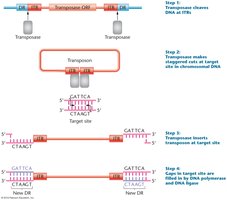
Transposon Insertion and Target Site Duplication
Transposase cleaves DNA at ITRs, makes staggered cuts at the target site, inserts the transposon, and fills gaps with DNA polymerase and ligase, creating new DRs.
Staggered cuts: Allow for integration and duplication of target site sequences.
Gap filling: DNA polymerase and ligase fill in gaps, completing the insertion.
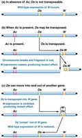
Ac-Ds System in Maize
The Ac-Ds system in maize demonstrates the interaction between autonomous (Ac) and nonautonomous (Ds) elements. Ac produces transposase, enabling Ds to move.
Ac (Activator): Autonomous, can transpose by itself.
Ds (Dissociation): Nonautonomous, requires Ac for movement.
Mutations: Ds insertion can disrupt gene function, leading to colorless kernels; excision restores function, causing spotted kernels.
Retrotransposons
Types of Retrotransposons
Retrotransposons are TEs that move via an RNA intermediate. They are classified as LTR (long terminal repeat) and non-LTR retrotransposons.
LTR retrotransposons: Contain long terminal repeats and encode integrase and reverse transcriptase.
Non-LTR retrotransposons: Lack LTRs but encode similar enzymes.
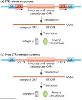
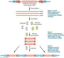
Mechanism of Retrotransposition
Retrotransposons are transcribed into RNA, translated to produce reverse transcriptase and integrase, and then reverse transcribed into DNA, which is inserted into the genome.
Transcription: RNA polymerase transcribes the retrotransposon.
Translation: Ribosomes produce reverse transcriptase and integrase.
Reverse transcription: RNA is converted to DNA.
Insertion: Integrase inserts the DNA into a new genomic location.
Copia Elements in Drosophila
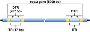
Structure and Function of Copia Elements
Copia elements are a class of retrotransposons in Drosophila, characterized by direct terminal repeats (DTRs) and inverted terminal repeats (ITRs).
DTR: Direct terminal repeat, 267 bp long.
ITR: Inverted terminal repeat, 17 bp long.
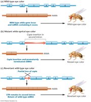
Effects of Copia Insertion
Copia insertion can disrupt gene function, such as altering eye color in Drosophila. Reversion can restore wild-type phenotype.
Mutation: Copia insertion in an intron can cause mutant phenotype.
Reversion: Partial loss of copia restores wild-type function.
Regulation of Gene Expression in Eukaryotes
Levels of Regulation and Chromatin Remodeling
Gene expression in eukaryotes is regulated at multiple levels, including chromatin structure, transcription, RNA processing, translation, and post-translational modification.
Chromatin remodeling: Alters accessibility of DNA to transcription machinery.
Histone modification: Covalent addition of functional groups to histone tails affects transcription.
Chromatin Structure and Histone Modification
Chromatin is composed of DNA wrapped around histone octamers, forming nucleosomes. Modification of histones (acetylation, methylation, phosphorylation) regulates gene expression.
Acetylation: Associated with increased transcription.
Methylation: Often suppresses transcription.
Phosphorylation: Can affect chromatin structure and gene expression.
Chromatin Remodeling Complexes
SWI/SNF is a well-studied chromatin remodeling complex that loosens DNA-histone interactions, making DNA accessible for transcription.
Repositioning: Nucleosomes are moved or removed to expose regulatory regions.
Gene activation: Facilitates binding of transcription factors and RNA polymerase II.
DNA Methylation and Genetic Imprinting
Direct methylation of DNA, especially cytosine residues, is used to turn genes off. Methyl groups recruit proteins that remove acetyl groups and block transcription factors.
Imprinting: Only one allele (based on parent sex) is expressed.
Regulation of Transcription
Structure of Eukaryotic Genes
Eukaryotic genes contain coding regions, promoters, enhancers, silencers, polyadenylation sites, and terminal regions. Exons are translated, while introns are not. 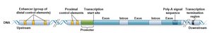
Cis-Acting Sequences
Cis-acting sequences are located on the same chromosome as the gene they regulate. They include promoters, enhancers, and silencers.
Promoters: On/off switch for transcription.
Enhancers: Regulate expression in specific tissues and levels.
Silencers: Repress gene expression.
Promoter Structure and Diversity
Promoters are composed of several elements, including the initiator (Inr), TATA box, TFIIB recognition element (BRE), downstream promoter element (DPE), and motif ten element (MTE). 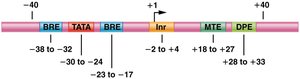
Proximal-Promoter Elements
Proximal-promoter elements, such as CAAT and GC boxes, are located upstream of the TATA and BRE motifs and enhance basal transcription levels. 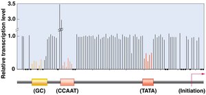
Enhancers and Silencers
Enhancers and silencers are cis-acting elements that regulate transcription initiation. Enhancers can be located far from the gene, while silencers repress transcription.
Transcription Factors
Transcription factors are proteins that bind to cis-acting sites and regulate gene expression. Activators increase transcription, while repressors decrease it.
DNA-binding domain: Binds to specific DNA sequences.
Trans-activating domain: Interacts with other proteins to modulate transcription.
Formation of RNA Pol II Initiation Complex
General transcription factors assemble at the promoter to form the pre-initiation complex (PIC), providing a platform for RNA polymerase II to recognize transcription start sites.
TFIID: Contains TATA binding protein (TBP) and TBP-associated factors (TAFs).
Additional factors: IIA, IIB, IIE, IIF, IIH.
Coactivators and Enhanceosome
Coactivators enable activators to contact promoter-bound factors, forming the enhanceosome, which interacts with the transcription complex to regulate transcription.
Posttranscriptional Regulation
Alternative Splicing
Alternative splicing generates different forms of mRNA from identical pre-mRNA, increasing the number of proteins produced from a single gene. This process is facilitated by small nuclear ribonucleoprotein particles (snRNPs).
Exons: Coding sequences that exit the nucleus.
Introns: Non-coding sequences removed during splicing.
Example: Calcitonin Gene
The calcitonin gene produces two distinct polypeptides via alternative splicing: calcitonin in the thyroid and calcitonin gene-related peptide (CGRP) in neurons.
Dscam Gene in Drosophila
The Dscam gene region contains highly variable exons that can be combined into over 38,000 unique polypeptides, guiding axon connections.
Control of mRNA Stability
The steady-state level of mRNA is determined by transcription and degradation rates. mRNA degradation involves shortening the poly-A tail, decapping, and endonuclease cleavage.
Protein Degradation: Ubiquitin and Proteosome
Ubiquitin tags proteins for degradation by the proteosome, a cylindrical structure that recycles amino acids.
p53 Protein Regulation
p53 is a transcription factor whose levels increase in response to DNA damage or stress. It activates transcription of the Mdm2 gene, which encodes a ubiquitin ligase that targets p53 for degradation.
Phosphorylation: Protects p53 from Mdm2-mediated degradation.
Cell survival signals: Activate Mdm2, leading to p53 degradation.
