 Back
BackLarge-Scale Chromosomal Changes: Structure, Variation, and Consequences
Study Guide - Smart Notes
 Tailored notes based on your materials, expanded with key definitions, examples, and context.
Large-Scale Chromosomal Changes
Introduction to Chromosome Structure and Variation
Large-scale chromosomal changes involve alterations in chromosome number or structure, which can have significant effects on phenotype, development, and evolution. Understanding these changes is essential for interpreting genetic disorders, evolutionary processes, and the mechanisms of heredity.
Chromosome Structure and Classification
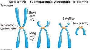
Chromosome Shapes and Features
Metacentric chromosomes: Centromere is in the middle, resulting in arms of equal length.
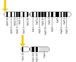
Submetacentric chromosomes: Centromere is slightly off-center, producing a short (p) and long (q) arm.
Acrocentric chromosomes: Centromere is near one end, creating a very short p arm and a satellite.
Telocentric chromosomes: Centromere is at the end, so there is no p arm.
Example: Human chromosomes display all types except telocentric.
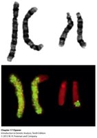
Visualizing Chromosomes
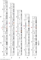
Giemsa (G) banding: Produces characteristic banding patterns for each chromosome, allowing identification and detection of structural changes.
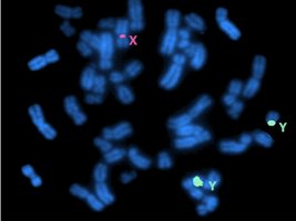
Fluorescence in situ hybridization (FISH): Uses fluorescent probes to label specific DNA sequences, enabling visualization of individual chromosomes or gene locations.
Multiplex FISH (m-FISH): Uses combinations of dyes to uniquely label all chromosomes in a karyotype.
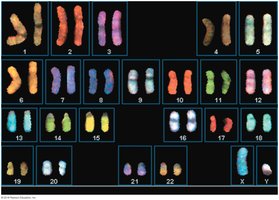
Example: m-FISH can distinguish all 24 human chromosomes by color.
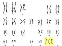
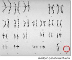
Standardization of Human Karyotypes
Human chromosomes are arranged in a standardized order for karyotyping, facilitating the detection of abnormalities.
Banding patterns are used to identify structural changes such as deletions, duplications, or translocations.
Chromosome Number Variations
Aneuploidy
Aneuploidy refers to the presence of an abnormal number of a particular chromosome, rather than a whole set. It is a common cause of genetic disorders in humans.
Monosomy (2n-1): Loss of a single chromosome.
Trisomy (2n+1): Gain of a single chromosome.

Example: Down syndrome (trisomy 21), Turner syndrome (monosomy X).
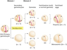
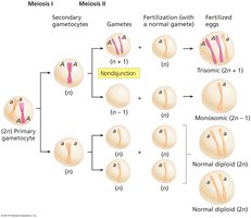
Mechanisms of Aneuploidy
Nondisjunction: Failure of homologous chromosomes or sister chromatids to separate properly during meiosis, resulting in gametes with abnormal chromosome numbers.
Fertilization: Fusion of an abnormal gamete with a normal one produces aneuploid zygotes.
Human Aneuploidy Syndromes
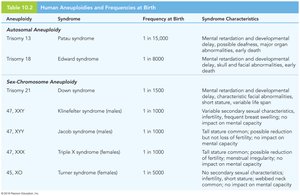
Most human aneuploidies are incompatible with life, but some result in viable syndromes with characteristic phenotypes.
Aneuploidy | Syndrome | Frequency at Birth | Syndrome Characteristics |
|---|---|---|---|
Trisomy 21 | Down syndrome | 1 in 1500 | Mental retardation, developmental delay, characteristic facial features, heart defects |
47, XXY | Klinefelter syndrome (male) | 1 in 1000 | Tall stature, infertility, mild cognitive impairment |
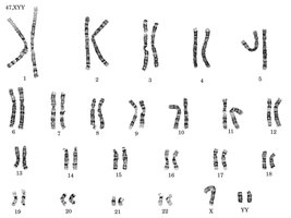
47, XYY | Jacob syndrome (male) | 1 in 1000 | Tall stature, normal fertility, possible learning difficulties |
47, XXX | Triple X syndrome (female) | 1 in 1000 | Tall stature, normal fertility, no major impact |
45, X | Turner syndrome (female) | 1 in 5000 | Short stature, infertility, webbed neck, normal intelligence |
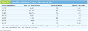
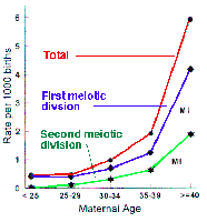
Maternal Age and Aneuploidy Risk
The risk of nondisjunction, especially for trisomy 21 (Down syndrome), increases with maternal age.
Meiosis I nondisjunction is more common than meiosis II nondisjunction at all ages.
Sex Chromosome Aneuploidy
Klinefelter, Jacob, Triple X, and Turner Syndromes
Klinefelter syndrome (XXY): Males with an extra X chromosome; tall, infertile, mild cognitive impairment.
Jacob syndrome (XYY): Males with an extra Y chromosome; tall, normal fertility, possible learning difficulties.
Triple X syndrome (XXX): Females with an extra X chromosome; tall, normal fertility, usually no major effects.
Turner syndrome (X0): Females missing one X chromosome; short stature, infertility, webbed neck.

Genetic Basis of Phenotypes in Sex Chromosome Aneuploidy
Haploinsufficiency: Some phenotypes, such as short stature in Turner syndrome, are due to insufficient dosage of genes like SHOX present on both X and Y chromosomes.
Chromosomal Mosaicism and Uniparental Disomy
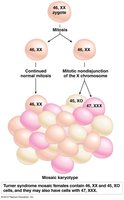
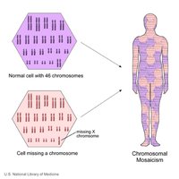
Chromosomal Mosaicism
Chromosomal mosaicism arises when mitotic nondisjunction occurs early in embryogenesis, resulting in an individual with two or more genetically distinct cell lines.
Accounts for 25-30% of Turner syndrome cases.
Can result in 45,X/46,XX or 45,X/46,XY karyotypes.
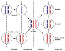
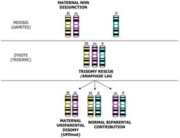
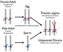
Uniparental Disomy (UPD)
UPD occurs when both copies of a chromosome are inherited from one parent. This can result from trisomy rescue and can lead to disorders if imprinted genes are involved.
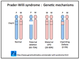
Prader-Willi syndrome: Loss of paternal genes on chromosome 15q leads to developmental and metabolic abnormalities.
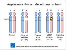
Angelman syndrome: Loss of maternal genes on chromosome 15q leads to neurological and developmental defects.
Epigenetics and Imprinting
Correct imprinting of genes on maternal or paternal chromosomes is essential for normal development. Imprinting disorders can result in syndromes such as Prader-Willi and Angelman.
Imprinting: Epigenetic silencing of one parental allele, so only the other is expressed.
Loss of the active allele (by deletion or UPD) leads to disease.
Euploidy and Polyploidy
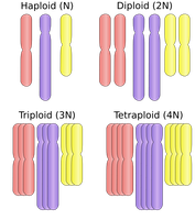
Definitions and Types
Euploidy: Variation in the number of complete sets of chromosomes (e.g., diploid, triploid, tetraploid).
Polyploidy: More than two sets of chromosomes; common in plants, rare in animals.
Aneuploidy: Variation in the number of individual chromosomes, not whole sets.
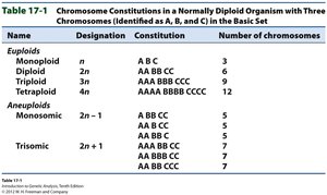
Name | Designation | Constitution | Number of chromosomes |
|---|---|---|---|
Monoploid | n | A B C | 3 |
Diploid | 2n | AA BB CC | 6 |
Triploid | 3n | AAA BBB CCC | 9 |
Tetraploid | 4n | AAAA BBBB CCCC | 12 |
Monosomic | 2n-1 | AA BB C | 5 |
Trisomic | 2n+1 | AA BB CCC | 7 |
Types of Polyploidy
Autopolyploidy: Multiple chromosome sets from the same species (e.g., autotetraploid potatoes).
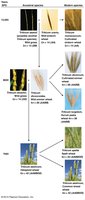
Allopolyploidy: Chromosome sets from different species (e.g., triticale, a wheat-rye hybrid).
Polyploidy in Nature and Agriculture
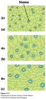
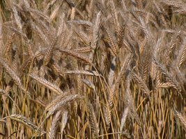
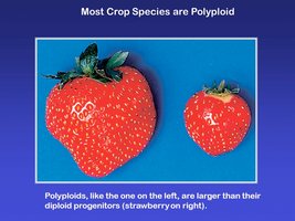
Polyploidy is common in plants and has played a major role in the evolution of crops.
Polyploids often have larger cells and organs, leading to larger fruits or flowers.
Consequences of Polyploidy
Odd-numbered polyploids (e.g., triploids) are usually sterile due to irregular chromosome segregation during meiosis.
Even-numbered polyploids (e.g., tetraploids) can produce functional gametes.
Structural Chromosome Changes
Deletions and Duplications
Terminal deletion: Loss of genes at the end of a chromosome.
Interstitial deletion: Loss of an internal segment of a chromosome.
Duplication: Repetition of a chromosome segment; can result in gene dosage effects or new gene functions.
Detection and Mapping of Deletions/Duplications
Deletions and duplications can be detected by banding analysis or FISH.
Deletion mapping uses pseudodominance to locate genes.
Inversions
Paracentric inversion: Does not include the centromere.
Pericentric inversion: Includes the centromere.
Inversion loops form during meiosis in inversion heterozygotes, leading to abnormal crossover products.
Translocations
Reciprocal translocation: Exchange of segments between two nonhomologous chromosomes.
Robertsonian translocation: Fusion of two acrocentric chromosomes, reducing chromosome number.
Translocation heterozygotes form cross-shaped structures during meiosis.
Clinical and Evolutionary Implications
Chromosome Abnormalities and Human Health
Chromosome abnormalities account for nearly 50% of spontaneous abortions.
Many structural and numerical changes are incompatible with life or cause severe syndromes.
Evolutionary Role of Chromosomal Changes
Polyploidy and structural rearrangements have driven the evolution of new species, especially in plants.
Hybridization and chromosome doubling can create fertile new species (e.g., wheat, triticale).
Additional info: Chromosomal changes are also important in cancer genetics, where rearrangements can activate oncogenes or inactivate tumor suppressors.
