 Back
BackMitosis and Cell Structure: Foundations for Genetic Continuity
Study Guide - Smart Notes
 Tailored notes based on your materials, expanded with key definitions, examples, and context.
Cell Structure and Genetic Function
Overview of Cell Types
Cells are the fundamental units of life, and their structure is closely tied to their genetic function. There are two main types of cells: prokaryotic (bacteria, archaea) and eukaryotic (plants, fungi, animals). All cells share common features such as a plasma membrane, DNA, and ribosomes.
Prokaryotic cells: Unicellular, lack a membrane-bound nucleus, DNA is found in a region called the nucleoid.
Eukaryotic cells: Typically multicellular, possess a true nucleus and membrane-bound organelles.

Plasma Membrane and Glycocalyx
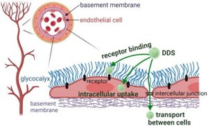
The plasma membrane surrounds all cells, separating them from the external environment and regulating the movement of substances in and out. In animal cells, the glycocalyx (cell coat) covers the plasma membrane and contains receptors for signal detection, aiding in cell communication and response to environmental cues.
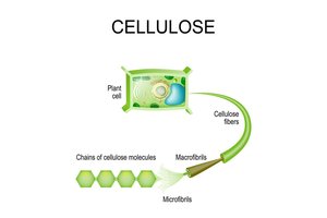
Plant cells: Have a cell wall made mostly of cellulose, providing structural support.
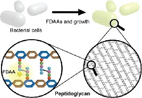
Bacterial cells: Cell walls contain peptidoglycan, a polymer that provides rigidity.
Cytoplasm and Cytoskeleton
The cytoplasm is a jelly-like substance inside the cell that contains organelles and is the site of many metabolic activities. The cytoskeleton is a network of fibers (microtubules and microfilaments) that provides structural support and facilitates cell movement.
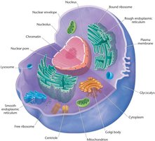
Mitochondria, Chloroplasts, and Endoplasmic Reticulum
Mitochondria are the sites of oxidative respiration and ATP production in both animal and plant cells. Chloroplasts (in plants and algae) are the sites of photosynthesis. The endoplasmic reticulum (ER) compartmentalizes the cytoplasm and increases surface area for biochemical synthesis:
Smooth ER (SER): Site of fatty acid and phospholipid synthesis.
Rough ER (RER): Studded with ribosomes; site of protein synthesis.
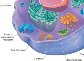
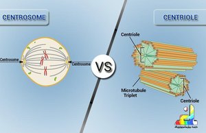
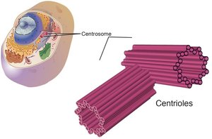
Centrioles and Centrosomes
Centrioles are cylindrical organelles found in the centrosome of animal cells. They organize spindle fibers for chromosome movement during mitosis and meiosis. Centrosomes are the microtubule-organizing centers of the cell.
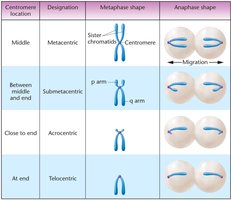
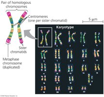
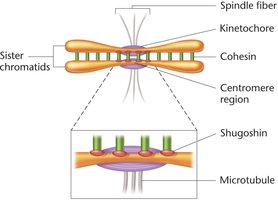
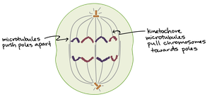
Centromeres and Chromosome Structure
The centromere is a constricted region on a chromosome that determines its shape and is the attachment site for spindle fibers via kinetochores. Chromosomes are classified based on centromere position:
Centromere Location | Designation | Metaphase Shape | Anaphase Shape |
|---|---|---|---|
Middle | Metacentric | \(\text{Sister chromatids with centromere in the middle}\) | \(\text{Migration to poles}\) |
Between middle and end | Submetacentric | \(p\) arm (short), \(q\) arm (long) | \(\text{Migration to poles}\) |
Close to end | Acrocentric | \(\text{Centromere near end}\) | \(\text{Migration to poles}\) |
At end | Telocentric | \(\text{Centromere at end}\) | \(\text{Migration to poles}\) |
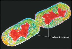
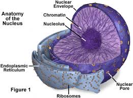
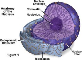
Nucleus and Nucleoid
The nucleus is a membrane-bound structure in eukaryotes that houses genetic material (DNA) organized as chromatin. It contains the nucleolus, the site of ribosomal RNA synthesis. In prokaryotes, the nucleoid is a non-membrane-bound region containing circular DNA.
Nucleus: Membrane-bound, linear DNA with histones (eukaryotes).
Nucleoid: Not membrane-bound, circular DNA, usually without histones (prokaryotes).
Genetic Material Organization
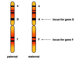
DNA contains the genetic instructions for growth, function, and response to stimuli. The genome is all the DNA in an organism. A gene is a segment of DNA that codes for a specific protein, and a locus is the location of a gene on a chromosome.
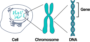
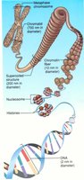
DNA Packaging
DNA is wrapped around histone proteins to form nucleosomes, which coil to form chromatin. Chromatin further condenses to form chromosomes during cell division.
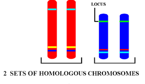
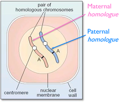
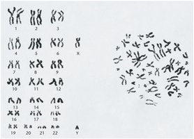
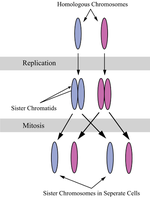
Chromosome Number and Homologous Chromosomes
Each eukaryotic species has a characteristic number of chromosomes. In humans, somatic cells have 46 chromosomes (23 pairs), while gametes have 23 chromosomes. Chromosomes exist as homologous pairs, with one chromosome from each parent. Homologous chromosomes carry genes for the same traits but may have different alleles.

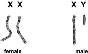
Sex Chromosomes and Autosomes
In humans, there are 22 pairs of autosomes and one pair of sex chromosomes (X and Y). Sex chromosomes determine biological sex and are not homologous in males (XY).
Mitosis: Partitioning Chromosomes into Dividing Cells
Overview of the Eukaryotic Cell Cycle
The cell cycle is the life of a cell from its formation to its division. It consists of interphase (cell growth and DNA replication) and the mitotic (M) phase (mitosis and cytokinesis).
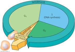
Interphase: G1 (cell growth), S (DNA synthesis/replication), G2 (final preparations for mitosis).
Mitotic phase: Mitosis (nuclear division) and cytokinesis (cytoplasmic division).
Stages of Mitosis
Mitosis is the process by which a cell divides its nucleus and genetic material to produce two genetically identical daughter cells. It consists of five stages:
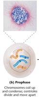
Prophase: Chromatin condenses into chromosomes, nuclear envelope disassembles, spindle fibers form, centrioles move to poles.
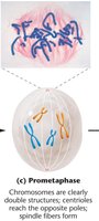
Prometaphase: Nuclear envelope fragments, spindle fibers attach to kinetochores on chromosomes.
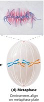
Metaphase: Chromosomes align at the metaphase plate, spindle fibers fully attached.
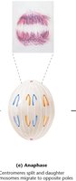
Anaphase: Sister chromatids separate at the centromere and move to opposite poles.
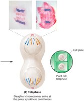
Telophase: Chromosomes decondense, nuclear envelopes reform, cytokinesis divides the cytoplasm.

Prophase and Sister Chromatids
During prophase, each chromosome consists of two identical sister chromatids joined at the centromere. The mitotic spindle begins to form, and the nuclear envelope breaks down.
Prometaphase
In prometaphase, the nuclear envelope is fully disassembled, and spindle fibers attach to kinetochores on the centromeres of chromosomes.
Metaphase
During metaphase, chromosomes align along the metaphase plate. This stage is ideal for karyotyping, as chromosomes are most condensed and visible.
Anaphase
In anaphase, sister chromatids are separated at the centromere and pulled to opposite poles by spindle fibers. Once separated, each chromatid is considered a daughter chromosome.
Telophase and Cytokinesis
During telophase, chromosomes decondense, nuclear envelopes reform, and the spindle apparatus disassembles. Cytokinesis divides the cytoplasm, resulting in two genetically identical daughter cells. In plant cells, a cell plate forms; in animal cells, a cleavage furrow forms.
Cell Cycle Regulation and Checkpoints
The cell cycle is tightly regulated by checkpoints at G1, S, G2, and M phases. These checkpoints ensure that the cell is ready to proceed to the next stage and help prevent errors in cell division. Cdc genes and cyclins produce proteins that regulate the cycle. If errors are detected, the cycle is halted for repair.
Summary
Mitosis is essential for growth, development, and tissue repair in multicellular organisms. It ensures genetic continuity by producing two identical daughter cells. The process is highly regulated to maintain genomic integrity.
