 Back
BackMitosis and Meiosis: Cell Structure, Chromosome Behavior, and Genetic Variation
Study Guide - Smart Notes
 Tailored notes based on your materials, expanded with key definitions, examples, and context.
Mitosis and Meiosis
Introduction to Cell Structure and Genetic Function
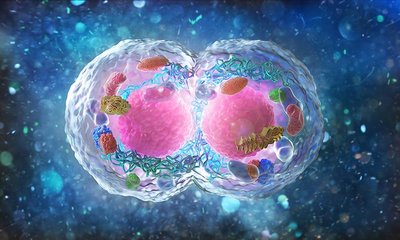
Cell structure is fundamentally linked to genetic function, as the organization and compartmentalization of cellular components facilitate the transmission and expression of genetic material. Eukaryotic cells transmit genetic material via chromosomes through mitosis and meiosis, processes essential for growth, development, and reproduction.
Genetic Material: DNA organized into chromosomes; viruses are excluded from this definition.
Major Processes: Mitosis produces two diploid somatic cells; Meiosis produces haploid gametes or spores.
Chromatin: During nondivisional phases, chromosomes uncoil into chromatin, a diffuse network within the nucleus.
Cell Types and Common Features
Cells are classified as prokaryotic or eukaryotic, each with distinct structural features but sharing fundamental components.
Prokaryotic Cells: Bacteria and archaea; lack membrane-bound organelles.
Eukaryotic Cells: Protists, plants, fungi, animals; contain membrane-bound organelles.
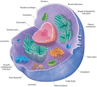
Common Features: Plasma membrane, DNA, ribosomes.
Plasma Membrane and Cell Wall
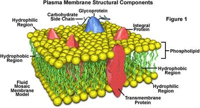
The plasma membrane surrounds all cells, delimiting them from the external environment. Plant cells also possess a cell wall composed mainly of cellulose, while bacterial cell walls contain peptidoglycan.
Glycocalyx: A glycoprotein and polysaccharide covering on animal cells, providing biochemical identity and receptor sites for signal transduction.

Nucleus and Nucleoid
The nucleus is a membrane-bound organelle in eukaryotes, housing DNA and the nucleolus, where ribosomal RNA is synthesized. In prokaryotes, the nucleoid is a non-membrane-bound region containing DNA.
Cytoplasm and Cytoskeleton
The cytoplasm contains organelles and is supported by the cytoskeleton, an extensive network of tubules and filaments.
Microtubules: Composed of tubulin; provide structural support and facilitate chromosome movement.
Microfilaments: Derived from actin; involved in cell shape and movement.

Endoplasmic Reticulum
The endoplasmic reticulum (ER) compartmentalizes the cytoplasm and increases surface area for biochemical synthesis.
Smooth ER (SER): Site of fatty acid and phospholipid synthesis.
Rough ER (RER): Studded with ribosomes; site of protein synthesis.

Mitochondria and Chloroplasts
Mitochondria and chloroplasts are cytoplasmic organelles responsible for energy production and photosynthesis, respectively. Both contain extranuclear DNA and can duplicate, transcribe, and translate their own genetic information.
Mitochondria: Site of oxidative phases of cell respiration, generating ATP.
Chloroplasts: Site of photosynthesis in plants, algae, and protozoans.

Centrioles
Centrioles are found in the centrosome of animal and plant cells and organize spindle fibers for chromosome movement during mitosis and meiosis.

Chromosomes in Diploid Organisms
Homologous Chromosomes
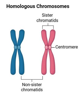
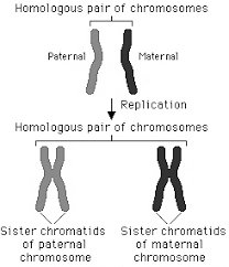
Chromosomes exist in homologous pairs in diploid organisms. Homologous chromosomes carry genes for the same inherited characteristics but may have different alleles.
Alleles: Alternative forms of the same gene.
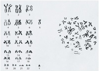
Somatic Cells: Humans have 46 chromosomes (23 pairs), representing the diploid number (2n).

Centromere and Chromosome Classification
The centromere is a constricted region on chromosomes, and its location determines chromosome appearance and classification.
Centromere Location | Designation | Metaphase Shape | Anaphase Shape |
|---|---|---|---|
Middle | Metacentric | Sister chromatids with centromere | Migration to poles |
Between middle and end | Submetacentric | p arm (short), q arm (long) | Migration to poles |
Close to end | Acrocentric | Centromere near end | Migration to poles |
At end | Telocentric | Centromere at terminal end | Migration to poles |
Karyotype and Genome
A karyotype illustrates the physical appearance of homologous chromosome pairs. The genome refers to the genetic information in a haploid set.
Biparental Inheritance and Alleles
Diploid organisms inherit genes from both parents, resulting in two copies of each gene. Alleles are alternative forms of the same gene, and each gene site is called a locus.

Sex-Determining Chromosomes
Sex chromosomes (e.g., X and Y in humans) are not homologous but behave as homologs during meiosis.
Mitosis: Partitioning Chromosomes
Cell Cycle and Mitosis
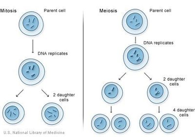
Mitosis partitions chromosomes into dividing cells, producing daughter cells with a full diploid complement. The cell cycle consists of interphase and mitosis.
Interphase: Includes S phase (DNA synthesis) and gap phases (G1, G2).
G0 Phase: Nondividing, metabolically active state.
Stages of Mitosis
Mitosis is divided into discrete stages: prophase, prometaphase, metaphase, anaphase, and telophase.
Prophase: Centrioles divide and move to poles; nuclear envelope breaks down; chromosomes condense.
Prometaphase: Chromosomes move to the equatorial plane (metaphase plate); spindle fibers form.
Metaphase: Chromosomes align on the metaphase plate; spindle fibers attach to kinetochores.
Anaphase: Sister chromatids separate and migrate to opposite poles.
Telophase: Chromosomes uncoil; nuclear envelope reforms; cytokinesis produces two new cells.
Cell Cycle Regulation and Checkpoints
Cell cycle regulation is controlled by cyclins and cyclin-dependent kinases (CDKs), which ensure proper progression and monitor for errors.
CDC Mutations: Affect enzymes called kinases, which regulate the cell cycle.
Checkpoints: Monitor mitosis for errors.
Meiosis: Creation of Haploid Gametes and Genetic Variation
Meiosis Overview
Meiosis reduces the amount of genetic material by half, producing haploid gametes or spores. It consists of two divisions: Meiosis I (reductional) and Meiosis II (equational).
Crossing Over: Genetic exchange between homologous chromosomes during prophase I, increasing genetic variation.
Interkinesis: Occurs between Meiosis I and II.
Meiosis I: Prophase I Substages
Leptonema: Chromosomes appear as long, single threads.
Zygonema: Synapsis occurs; homologous chromosomes pair as bivalents.
Pachynema: Bivalents become tetrads; crossing over occurs.
Diplonema: Sister chromatids separate within tetrads; chiasmata form.
Diakinesis: Nuclear envelope breaks down; centromeres attach to spindle fibers.
Meiosis I: Metaphase I, Anaphase I, Telophase I
Metaphase I: Chromosomes at maximum shortness; terminal chiasmata hold nonsister chromatids together.
Anaphase I: Homologous chromosomes separate; nondisjunction may occur.
Telophase I: Cytokinesis produces two haploid cells; nuclear membranes form.
Meiosis II: Second Meiotic Division
Prophase II: Each dyad consists of sister chromatids.
Metaphase II: Centromeres align on the equatorial plate.
Anaphase II: Sister chromatids separate to opposite poles.
Telophase II: Each chromosome is now a monad; cytokinesis results in four haploid cells.
Development of Gametes: Spermatogenesis vs. Oogenesis
Spermatogenesis
Male gametes are produced in the testes. The primary spermatocyte undergoes meiosis I to produce two secondary spermatocytes, which undergo meiosis II to produce four haploid spermatids.
Oogenesis
Female gametes are produced in the ovary. Four daughter cells do not receive equal cytoplasm; only one becomes the ovum, while the others become polar bodies.
Meiosis and Sexual Reproduction
Importance of Meiosis
Meiosis is critical for reducing diploid genetic information to haploid, enabling successful sexual reproduction in all diploid organisms. Plant and fungi life cycles alternate between diploid and haploid stages.
Physical Nature of Mitotic and Meiotic Chromosomes
Chromosome Structure
Chromosomes are visible only during mitosis and meiosis, when chromatin fibers coil and condense. Electron microscopy has revealed the folded-fiber model of chromosome structure.
Case Study: Effects of Chemotherapy and Radiotherapy on Spermatogenesis
Clinical Implications
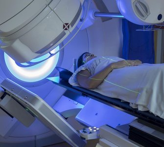
Intermittent chemotherapy and radiotherapy can temporarily reduce mature sperm count and cause abnormal chromosome numbers in developing spermatocytes. These abnormalities typically resolve within 40-74 days post-treatment, highlighting the sensitivity of spermatogenesis to environmental factors.
Summary Table: Haploid Numbers in Various Organisms
Common Name | Scientific Name | Haploid Number |
|---|---|---|
Black bread mold | Aspergillus nidulans | 8 |
Broad bean | Vicia faba | 6 |
Chimpanzee | Pan troglodytes | 24 |
Corn | Zea mays | 10 |
Cotton | Gossypium hirsutum | 26 |
Dog | Canis familiaris | 39 |
Fruit fly | Drosophila melanogaster | 4 |
Garden pea | Pisum sativum | 7 |
House mouse | Mus musculus | 20 |
Human | Homo sapiens | 23 |
Jimson weed | Datura stramonium | 12 |
Pink bread mold | Neurospora crassa | 7 |
Roundworm | Caenorhabditis elegans | 6 |
Wheat | Triticum aestivum | 21 |
Yeast | Saccharomyces cerevisiae | 16 |
Zebrafish | Danio rerio | 25 |
Key Equations
Diploid Number:
Haploid Number:
Comparison Table: Mitosis vs. Meiosis
Process | Outcome | Chromosome Number | Genetic Variation |
|---|---|---|---|
Mitosis | 2 daughter cells | Diploid (2n) | Identical to parent |
Meiosis | 4 daughter cells | Haploid (n) | Genetically varied |
