 Back
BackMitosis and Meiosis: Cellular and Chromosomal Foundations
Study Guide - Smart Notes
 Tailored notes based on your materials, expanded with key definitions, examples, and context.
Mitosis and Meiosis: Cellular and Chromosomal Foundations
Introduction to Cell Types and Genetic Material
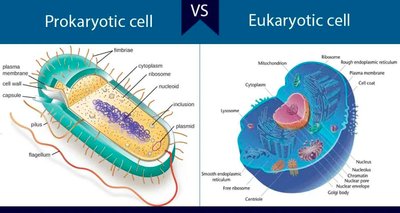
All living organisms are composed of cells, which can be classified as either prokaryotic or eukaryotic. Both cell types contain DNA, the hereditary material, and ribosomes for protein synthesis. Understanding the differences between these cell types is fundamental to genetics, as it underpins the mechanisms of inheritance and cell division.
Prokaryotic cells: Lack a nucleus; DNA is found in a nucleoid region.
Eukaryotic cells: Possess a membrane-bound nucleus containing DNA.
Common features: Plasma membrane, DNA, ribosomes.
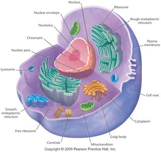
Eukaryotic Cell Structure
The nucleus is a defining feature of eukaryotic cells, serving as the repository for genetic material. Within the nucleus, DNA exists as chromatin when the cell is not dividing, and condenses into chromosomes during cell division.
Nucleus: Membrane-bound organelle containing DNA.
Chromatin: Granular form of DNA and proteins in the nucleus during interphase.
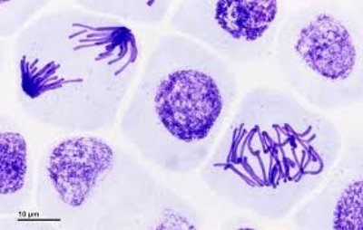
Chromosomes: Linear structures composed of DNA and proteins, visible during cell division.
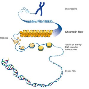
DNA Organization: Chromatin and Chromosomes
In eukaryotes, DNA is packaged with proteins (histones) to form chromatin, which further condenses into chromosomes during cell division. This hierarchical organization is crucial for the regulation of gene expression and the accurate segregation of genetic material.
Chromatin fiber: DNA wrapped around histones, forming nucleosomes.
Chromosome: Highly condensed chromatin visible during mitosis and meiosis.
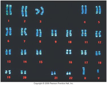
Chromosome Number and Ploidy
Cells are classified based on the number of chromosome sets they contain. Diploid cells have two sets of chromosomes, while haploid cells have one. The number of chromosomes varies widely among species and is a key factor in inheritance and genetic diversity.
Diploid (2N): Two sets of chromosomes (e.g., human somatic cells have 46 chromosomes).
Haploid (N): One set of chromosomes (e.g., human gametes have 23 chromosomes).
Species variation: Chromosome number differs among organisms (e.g., fruit fly: 8, dog: 78).
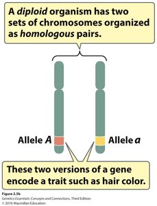
Homologous Chromosomes and Genetic Variation
In diploid organisms, chromosomes exist in homologous pairs. Each pair consists of chromosomes that carry genes for the same traits, but may have different versions (alleles) of those genes. This arrangement is fundamental to genetic inheritance and variation.
Homologous chromosomes: Similar in structure and gene content, but may carry different alleles.
Alleles: Different versions of a gene (e.g., allele A and allele a for hair color).
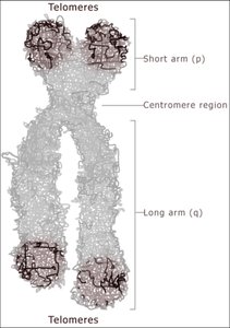
Sister Chromatids, Centromeres, and Telomeres
During cell division, each chromosome is replicated to form two sister chromatids, joined at a region called the centromere. The centromere is essential for proper chromosome segregation. Telomeres are repetitive DNA sequences at the ends of chromatids, protecting chromosomes from deterioration.
Sister chromatids: Identical copies of a chromosome, joined at the centromere.
Centromere: Constricted region linking sister chromatids; determines chromosome shape.
Telomeres: Protective sequences at chromosome ends.
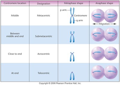
Centromere Placement and Chromosome Classification
Chromosomes are classified based on the position of the centromere, which affects their shape during cell division. The main types are metacentric, submetacentric, acrocentric, and telocentric.
Centromere location | Designation | Metaphase shape | Anaphase shape |
|---|---|---|---|
Middle | Metacentric | p arm, q arm, centromere in center | Equal migration of arms |
Between middle and end | Submetacentric | Centromere slightly off-center | Unequal migration |
Close to end | Acrocentric | Centromere near one end | One long, one short arm |
At end | Telocentric | Centromere at tip | Single arm migration |
Overview of Cell Division: Mitosis and Meiosis
Cell division is essential for growth, development, and reproduction. Mitosis produces genetically identical cells for growth and repair, while meiosis generates haploid gametes for sexual reproduction, introducing genetic diversity.
Mitosis: One division, produces two identical diploid cells.
Meiosis: Two divisions, produces four genetically unique haploid cells.
Key terms: Chromosome, chromatid, centromere, homologous pair, allele, ploidy.
Genetic condition changes: Diploid to haploid during gamete formation; restoration of diploidy at fertilization.
Additional info:
True or False: Sister chromatids can be heterozygous. False. Sister chromatids are identical copies formed during DNA replication; homologous chromosomes can be heterozygous.
Chromosome number is species-specific and does not correlate with organism complexity.
