 Back
BackCh. 2 Mitosis and Meiosis: Cellular Basis of Genetic Continuity
Study Guide - Smart Notes
 Tailored notes based on your materials, expanded with key definitions, examples, and context.
Chapter 2: Mitosis and Meiosis
Introduction to Genetic Continuity
Genetic continuity in eukaryotic organisms is maintained through two fundamental processes: mitosis and meiosis. These processes ensure the accurate transmission of genetic material from one generation to the next and are essential for growth, development, and reproduction.
DNA is the genetic material in all living organisms (except some viruses).
Genes are organized into chromosomes.
Mitosis produces genetically identical daughter cells (2n).
Meiosis produces gametes or spores with half the chromosome number (n), introducing genetic variation.
Cell Structure and Genetic Function
Prokaryotic vs. Eukaryotic Cells
Cells are classified as prokaryotic or eukaryotic based on their internal structure, which is closely tied to their genetic processes.
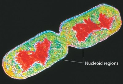
Prokaryotes: Lack a nucleus and membrane-bound organelles; genetic material is a single, circular DNA molecule in the nucleoid region.
Eukaryotes: Have a nucleus and membrane-bound organelles; DNA is organized with proteins (histones) into chromatin within the nucleus.
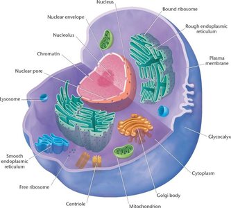
Key Cellular Components Involved in Genetics
Nucleus: Contains genetic material (DNA) and nucleolus (site of rRNA synthesis).
Ribosomes: Sites of protein synthesis, where mRNA is translated.
Mitochondria and Chloroplasts: Contain their own DNA, supporting the endosymbiotic theory.
Centrioles and Centrosome: Organize spindle fibers for chromosome movement during cell division.
Plasma Membrane: Defines cell boundary and regulates material movement.
Cell Wall (plants): Provides structural support, composed mainly of cellulose.
Chromosomes and Karyotypes
Chromosome Structure and Classification
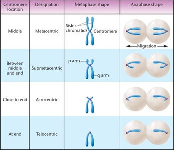
Chromosomes are classified based on the position of the centromere, which affects their shape and behavior during cell division.
Centromere Location | Designation | Metaphase Shape | Anaphase Shape |
|---|---|---|---|
Middle | Metacentric | V-shaped | Even migration |
Between middle and end | Submetacentric | J-shaped (p and q arms) | Uneven migration |
Close to end | Acrocentric | Very short p arm | Distinct migration |
At end | Telocentric | Rod-shaped | Linear migration |
Homologous Chromosomes and Karyotypes
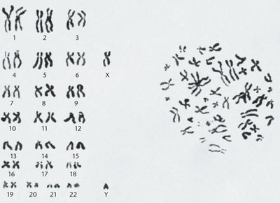
Somatic cells of diploid organisms contain pairs of homologous chromosomes, each carrying the same genes at corresponding loci. The karyotype is the complete set of chromosomes in a species, arranged and displayed for analysis.
Humans have 46 chromosomes (2n = 46), arranged in 23 pairs.
Homologous chromosomes carry alleles—alternative forms of the same gene.
Sex chromosomes (X and Y) are nonhomologous in males.
Haploid and Diploid Numbers
The haploid number (n) is half the diploid number (2n) and represents the number of unique chromosomes in a gamete. The genome is the complete set of genetic information in one haploid set.
Common Name | Scientific Name | Haploid Number (n) |
|---|---|---|
Human | Homo sapiens | 23 |
Fruit fly | Drosophila melanogaster | 4 |
Corn | Zea mays | 10 |
Chimpanzee | Pan troglodytes | 24 |
Yeast | Saccharomyces cerevisiae | 16 |
Mitosis: Partitioning Chromosomes
The Cell Cycle and Interphase
The cell cycle is the sequence of events in the life of a cell, alternating between division and non-division. It consists of interphase (G1, S, G2) and mitosis (M phase).
G1 phase: Cell growth and preparation for DNA synthesis.
S phase: DNA replication.
G2 phase: Preparation for mitosis; cell grows further.
G0 phase: Non-dividing, metabolically active state.
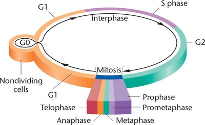
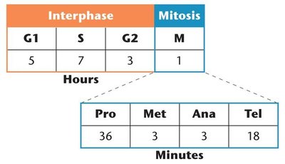
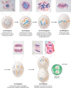
Stages of Mitosis
Mitosis is divided into distinct stages, each with specific events ensuring equal distribution of chromosomes.
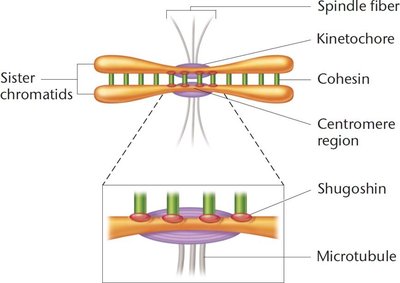
Prophase: Chromosomes condense, nuclear envelope breaks down, spindle apparatus forms.
Prometaphase: Chromosomes attach to spindle fibers via kinetochores and begin to move.
Metaphase: Chromosomes align at the metaphase plate.
Anaphase: Sister chromatids separate and move to opposite poles.
Telophase: Nuclear envelopes reform, chromosomes decondense, cytokinesis divides the cytoplasm.
Regulation of the Cell Cycle
The cell cycle is tightly regulated by kinases and cyclins. Checkpoints at G1, G2, and M phases ensure errors are corrected or the cell is removed from the cycle, preventing malignancy.
Kinases: Enzymes that control cell cycle progression.
Cyclins: Proteins that activate kinases at specific points.
Checkpoints: Control mechanisms that verify whether the processes at each phase have been accurately completed.
Meiosis: Formation of Gametes and Genetic Variation
Overview of Meiosis
Meiosis is a two-division process that reduces the chromosome number by half, producing haploid gametes or spores. It is essential for sexual reproduction and introduces genetic diversity.
Meiosis I: Reductional division (homologous chromosomes separate).
Meiosis II: Equational division (sister chromatids separate).
Each division includes prophase, metaphase, anaphase, and telophase stages.
Key Events in Meiosis I
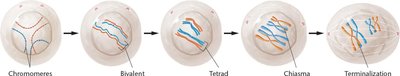
Prophase I: Homologous chromosomes pair (synapsis) and crossing over occurs, forming bivalents and tetrads.
Metaphase I: Tetrads align randomly at the metaphase plate; terminal chiasmata hold non-sister chromatids together.
Anaphase I: Homologous chromosomes (dyads) are separated to opposite poles (disjunction); nondisjunction can occur if separation fails.
Telophase I: Nuclear membranes reform around dyads; short interphase follows without DNA replication.


Meiosis II and Gamete Formation
Meiosis II resembles mitosis, where sister chromatids of each chromosome are separated, resulting in four haploid cells from one diploid cell.
Prophase II: Chromosomes condense again in each haploid cell.
Metaphase II: Chromosomes align at the metaphase plate.
Anaphase II: Centromeres divide, and sister chromatids move to opposite poles.
Telophase II: Nuclear membranes reform, and cytokinesis produces four genetically unique haploid gametes.
Genetic Variation: Crossing Over and Independent Assortment
Meiosis introduces genetic variation through two main mechanisms:
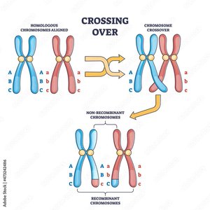
Crossing Over: Exchange of genetic material between non-sister chromatids during prophase I, resulting in recombinant chromosomes.
Independent Assortment: Random alignment of homologous pairs during metaphase I leads to different combinations of maternal and paternal chromosomes in gametes.
Significance of Meiosis in Sexual Reproduction
Meiosis is essential for the production of haploid gametes in animals and spores in plants, ensuring genetic continuity and diversity in diploid organisms. The reshuffling of alleles through crossing over and independent assortment increases genetic variability, which is fundamental to evolution and adaptation.
