 Back
BackMitosis and Meiosis: Cellular Basis of Genetic Inheritance
Study Guide - Smart Notes
 Tailored notes based on your materials, expanded with key definitions, examples, and context.
Chapter 2: Mitosis and Meiosis
Introduction
Genetic material in living organisms is composed of DNA, which is organized into chromosomes. In eukaryotes, the transmission of genetic material occurs through the processes of mitosis and meiosis. Mitosis produces genetically identical cells, while meiosis generates gametes or spores, introducing genetic variation.
2.1 Cell Structure Is Closely Tied to Genetic Function
Types of Cells
Prokaryotic cells: Include bacteria and archaea; lack a membrane-bound nucleus.
Eukaryotic cells: Include protists, plants, fungi, and animals; possess a membrane-bound nucleus and organelles.
All cells share common features: plasma membrane, DNA, and ribosomes.
Plasma Membrane and Cell Wall
The plasma membrane surrounds all cells, delimiting the cell from its environment.
Plant cells have a cell wall composed mainly of cellulose; bacterial cell walls contain peptidoglycan.
Glycocalyx
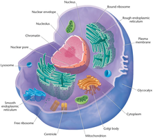
The glycocalyx (cell coat) is a covering on the plasma membrane of animal cells, made of glycoproteins and polysaccharides.
Functions in biochemical identity and cell signaling via receptor molecules.
Nucleus and Nucleoid
Nucleus: Found in eukaryotes, membrane-bound, houses DNA. Contains the nucleolus, where rRNA is synthesized.
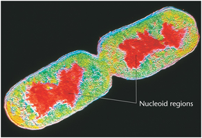
Nucleoid: Found in prokaryotes, not membrane-bound, contains genetic material.
Cytoplasm and Cytoskeleton
Cytoplasm: Includes organelles and cytosol (the colloidal material surrounding organelles).
Cytoskeleton: Network of microtubules (tubulin) and microfilaments (actin) providing structural support and facilitating movement.
Endoplasmic Reticulum (ER)
Rough ER (RER): Studded with ribosomes; site of protein synthesis.
Smooth ER (SER): Lacks ribosomes; site of fatty acid and phospholipid synthesis.
Mitochondria and Chloroplasts
Mitochondria: Site of oxidative phosphorylation and ATP production in both plant and animal cells.
Chloroplasts: Site of photosynthesis in plants, algae, and some protozoans.
Centrioles
Located in the centrosome of animal and some plant cells.
Organize spindle fibers for chromosome movement during cell division.
2.2 Chromosomes Exist in Homologous Pairs in Diploid Organisms
Homologous Chromosomes
Chromosomes in diploid organisms exist in homologous pairs, carrying genes for the same traits but possibly different alleles.
Humans have 46 chromosomes (23 pairs); the diploid number is denoted as 2n.
Each gene is located at a specific locus on a chromosome.
Centromere and Chromosome Morphology
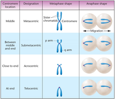
The centromere is a constricted region that determines chromosome shape and is essential for proper segregation during cell division.
Types of chromosomes based on centromere position:
Metacentric: Centromere in the middle
Submetacentric: Centromere between middle and end
Acrocentric: Centromere close to end
Telocentric: Centromere at the end
Centromere location | Designation | Metaphase shape | Anaphase shape |
|---|---|---|---|
Middle | Metacentric | Sister chromatids with centromere in the middle | Migration to poles |
Between middle and end | Submetacentric | p arm (short), q arm (long) | Migration to poles |
Close to end | Acrocentric | Centromere near end | Migration to poles |
At end | Telocentric | Centromere at end | Migration to poles |
Karyotype
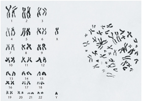
A karyotype is a visual representation of the complete set of chromosomes in a cell, arranged in homologous pairs.
Genome and Alleles
The genome is the complete set of genetic information in a haploid set of chromosomes.
Alleles are alternative forms of the same gene found at the same locus on homologous chromosomes.
Biparental inheritance: Diploid organisms inherit one chromosome of each pair from each parent.
Sex Chromosomes
Sex-determining chromosomes (e.g., X and Y in humans) are not homologous in size or genetic content but behave as homologs during meiosis.
2.3 Mitosis Partitions Chromosomes into Dividing Cells
Cell Cycle Overview
The cell cycle consists of interphase (G1, S, G2) and mitosis.
G0 phase: Nondividing but metabolically active state.
Karyokinesis: Division of the nucleus; cytokinesis: division of the cytoplasm.
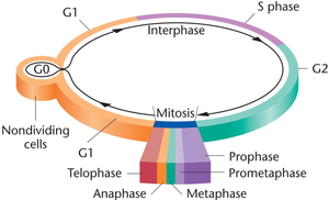
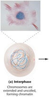
Interphase
During interphase, chromosomes are not visible and exist as chromatin.
DNA replication occurs in the S phase.
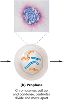
Mitosis Stages
Prophase: Chromosomes condense, centrioles move to poles, nuclear envelope breaks down.
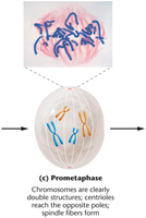
Prometaphase: Chromosomes move to the metaphase plate, spindle fibers form.
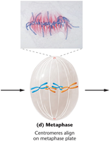
Metaphase: Chromosomes align at the metaphase plate; kinetochores attach to spindle fibers.
Anaphase: Sister chromatids separate and move to opposite poles, now called daughter chromosomes.
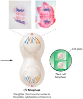
Telophase: Chromosomes decondense, nuclear envelope reforms, cytokinesis occurs.
Key Proteins in Chromosome Segregation
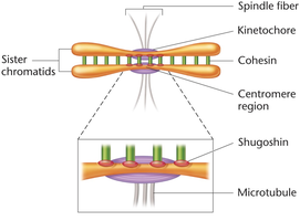
Cohesin: Protein complex holding sister chromatids together.
Separase: Enzyme that degrades cohesin to allow chromatid separation.
Shugoshin: Protects cohesin from premature degradation.
Cell Cycle Regulation
Regulated by cyclin-dependent kinases (CDKs) and cyclins.
Checkpoints monitor for errors and ensure proper division.
2.4 Meiosis Creates Haploid Gametes and Spores and Enhances Genetic Variation in Species
Overview of Meiosis
Meiosis reduces the chromosome number by half, producing haploid gametes or spores.
Consists of two sequential divisions: Meiosis I (reductional) and Meiosis II (equational).
DNA replication occurs only once, before Meiosis I.
Genetic Variation: Crossing Over
During prophase I, homologous chromosomes pair (synapsis) and exchange genetic material (crossing over) at chiasmata, increasing genetic diversity.
Stages of Meiosis
Each meiotic division includes prophase, metaphase, anaphase, and telophase.
Prophase I: Homologous chromosomes pair to form bivalents (tetrads); crossing over occurs.
Metaphase I: Homologous pairs align at the metaphase plate.
Anaphase I: Homologous chromosomes separate; sister chromatids remain attached.
Telophase I: Two haploid cells form; nuclear membranes may reform.
Meiosis II: Similar to mitosis; sister chromatids separate, resulting in four haploid cells.
Comparison: Mitosis vs. Meiosis
Mitosis: Produces two genetically identical diploid cells for growth and repair.
Meiosis: Produces four genetically unique haploid cells for sexual reproduction.
Summary
Mitosis and meiosis are essential for genetic continuity and variation.
Proper regulation and execution of these processes ensure healthy development and inheritance.
