 Back
BackMitosis: Mechanisms and Regulation of Somatic Cell Division
Study Guide - Smart Notes
 Tailored notes based on your materials, expanded with key definitions, examples, and context.
Mitosis and Cell Division
Overview of Mitosis
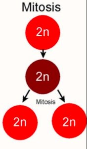
Mitosis is a fundamental process in eukaryotic cells that results in the production of two genetically identical somatic daughter cells. This process maintains the chromosome number and ploidy of the parent cell, ensuring genetic stability across cell generations. Mitosis is essential for growth, development, and tissue repair in multicellular organisms.
Somatic cells undergo mitosis to produce two identical daughter cells.
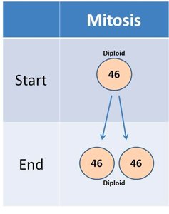
Each daughter cell receives the same number of chromosomes as the parent cell (diploid, 2n).
Mitosis does not generate genetic variation in daughter cells.
Comparison: Mitosis vs. Meiosis
While mitosis produces identical somatic cells, meiosis generates genetically distinct gametes or spores with half the chromosome number of the parent cell. This distinction is crucial for sexual reproduction and genetic diversity.
Mitosis: Maintains chromosome number (2n → 2n).
Meiosis: Reduces chromosome number by half (2n → n).
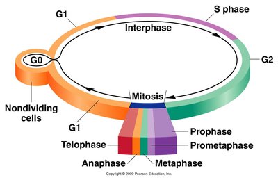
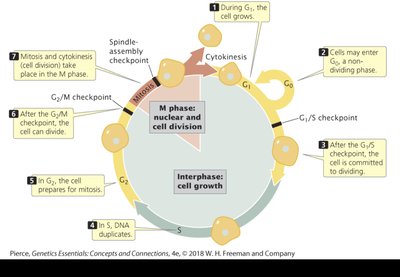
The Cell Cycle
Phases of the Cell Cycle
The cell cycle is a regulated sequence of events that includes cell growth, DNA replication, and cell division. It is divided into interphase (G1, S, G2) and the mitotic phase (M phase).
G1 phase: Cell grows and prepares for DNA synthesis.
S phase: DNA is replicated, resulting in sister chromatids.
G2 phase: Cell prepares for mitosis.
M phase: Includes nuclear division (karyokinesis) and cytoplasmic division (cytokinesis).
G0 phase: Nondividing, metabolically active state.
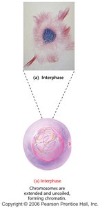
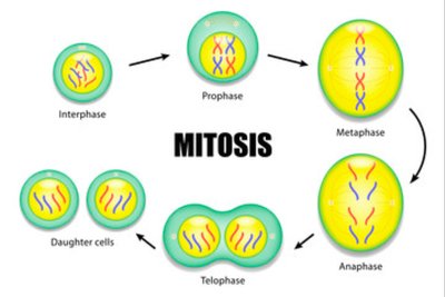
Interphase
Interphase is the period between mitotic divisions when the cell grows, replicates its DNA, and duplicates centrosomes. Chromosomes are relaxed and not easily visible under a light microscope.
Chromatin is the diffuse network of uncoiled chromosomes.
Centrosome duplication occurs in preparation for mitosis.
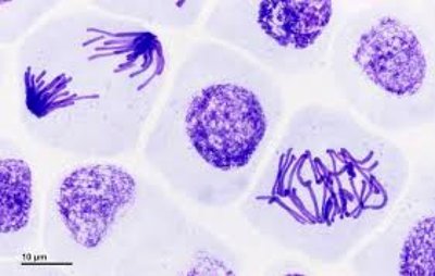
Stages of Mitosis
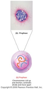
Prophase
During prophase, chromosomes condense and become visible, the nuclear envelope begins to break down, and centrosomes migrate to opposite poles, initiating spindle formation.
Chromosomes coil and shorten.
Centrosomes (composed of centrioles) move apart.
Spindle apparatus begins to form.
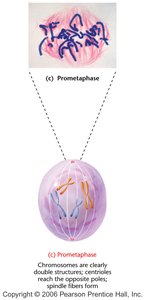
Prometaphase
In prometaphase, the nuclear envelope is fully disassembled, chromosomes are fully condensed, and spindle fibers attach to chromosomes via kinetochores. Chromosomes begin moving toward the metaphase plate.
Centrioles reach opposite poles.
Chromosomes attach to spindle fibers at kinetochores.
Metaphase
Chromosomes align at the metaphase plate, an imaginary plane equidistant from the spindle poles. This alignment ensures equal segregation of genetic material.
Centromeres align on the metaphase plate.
Kinetochores are attached to spindle microtubules.
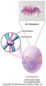
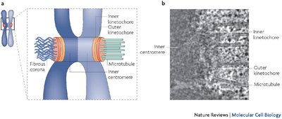
Centromere and Kinetochore Structure
The centromere is the region of the chromosome where sister chromatids are joined. The kinetochore is a protein complex at the centromere that attaches to spindle microtubules, facilitating chromosome movement during mitosis.
Centromere: DNA region joining sister chromatids.
Kinetochore: Protein structure for spindle attachment and chromatid movement.
Anaphase
During anaphase, sister chromatids separate (disjunction) and migrate toward opposite spindle poles, becoming individual daughter chromosomes.
Microtubule contraction at kinetochores pulls chromatids apart.
Ensures equal distribution of genetic material.
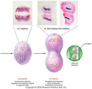
Telophase and Cytokinesis
Telophase marks the completion of chromosome migration, reformation of nuclear membranes, and relaxation of chromosomes. Cytokinesis divides the cytoplasm, resulting in two separate daughter cells.
Chromosomes decondense.
Nuclear envelopes reform.
Cytokinesis produces two genetically identical diploid cells.
Cell Cycle Checkpoints
Regulation of Mitosis
Cell cycle checkpoints are control mechanisms that ensure the fidelity of cell division. They monitor cell size, DNA integrity, and spindle attachment to prevent errors in mitosis.
G1/S checkpoint: Checks cell size and DNA condition.
G2/M checkpoint: Ensures DNA replication is complete and undamaged.
M checkpoint: Verifies spindle fiber attachment to kinetochores.
Results and Significance of Mitosis
Outcomes of Mitosis
Mitosis produces two daughter cells that are genetically identical to each other and to the parent cell, each with a full diploid complement of chromosomes. This process is crucial for organismal growth, tissue repair, and asexual reproduction.
Parent cell: Original cell before division.
Daughter cells: Genetically identical cells produced by mitosis.
No genetic variation is generated in mitosis.
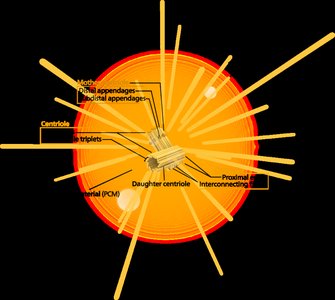
Key Structures in Mitosis
Centrosome
The centrosome is the main microtubule organizing center in animal cells, composed of centrioles. It plays a critical role in spindle formation and chromosome segregation during mitosis.
Duplicates during interphase.
Organizes spindle fibers for chromosome movement.
Summary Table: Mitosis vs. Meiosis
Process | Purpose | Chromosome Number | Genetic Variation |
|---|---|---|---|
Mitosis | Growth, repair, asexual reproduction | Maintained (2n → 2n) | No |
Meiosis | Sexual reproduction | Halved (2n → n) | Yes |
Review Questions
Where are the chromatids, centromeres, centrosomes, and spindle fibers during mitosis?
Can you tell whether the cells are diploid or haploid?
Does mitosis generate genetic variation in daughter cells? Why or why not?
Visual Summary: Mitosis in Action
Additional info: Mitosis is a highly regulated process, and errors in chromosome segregation can lead to aneuploidy and disease. Understanding the molecular mechanisms of mitosis is fundamental for genetics, cell biology, and medical research.
