 Back
BackMolecular Structure of DNA: Composition, Structure, and Forms
Study Guide - Smart Notes
 Tailored notes based on your materials, expanded with key definitions, examples, and context.
DNA and RNA Structure: Nucleotides as Building Blocks
Components of Nucleotides
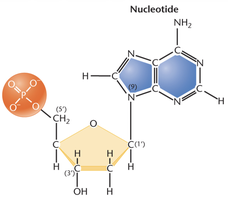
Nucleotides are the fundamental monomers that compose nucleic acids, DNA and RNA. Each nucleotide consists of three essential components:
Phosphate group
Pentose sugar (deoxyribose in DNA, ribose in RNA)
Nitrogenous base (purine or pyrimidine)
These nucleotides are linked together by phosphodiester bonds to form the sugar-phosphate backbone of nucleic acids, imparting directionality (5′ to 3′) to each strand.
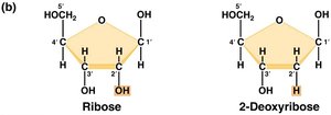
Comparison of Ribose and Deoxyribose
The pentose sugar distinguishes DNA from RNA. Ribose (in RNA) has a hydroxyl group (–OH) at the 2′ carbon, while deoxyribose (in DNA) lacks this oxygen atom, making DNA more chemically stable and suitable for long-term genetic storage.
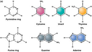
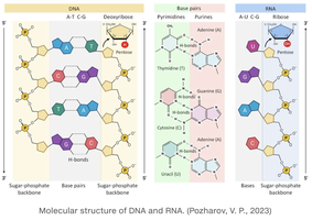
Nitrogenous Bases: Purines and Pyrimidines
Nitrogenous bases are categorized as either purines (adenine, guanine) or pyrimidines (cytosine, thymine, uracil). DNA contains adenine (A), guanine (G), cytosine (C), and thymine (T), while RNA contains uracil (U) instead of thymine.
Purines: Double-ring structures (A, G)
Pyrimidines: Single-ring structures (C, T, U)
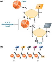
Formation of Nucleic Acid Polymers
Nucleotide Triphosphates and Phosphodiester Bonds
Nucleotide triphosphates (e.g., ATP, GTP) serve as precursors for nucleic acid synthesis. The energy released from the removal of terminal phosphate groups drives the formation of phosphodiester bonds between the 3′ hydroxyl of one nucleotide and the 5′ phosphate of the next, creating the sugar-phosphate backbone.
DNA Structure: Historical Evidence and Double Helix Model
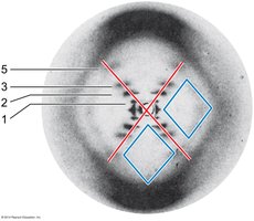
Franklin and Wilkins’ X-Ray Diffraction Evidence
X-ray diffraction studies by Rosalind Franklin and Maurice Wilkins provided the first direct evidence of DNA’s helical structure. The characteristic X-shaped diffraction pattern indicated a repeating helical structure with a periodicity of 3.4 Å per base pair and 10 base pairs per turn (34 Å per helical turn).
Watson and Crick’s Double Helix Model
Watson and Crick, using Franklin’s data and Chargaff’s base ratios, proposed the double helix model of DNA in 1953. The model features two antiparallel strands held together by complementary base pairing (A–T and G–C), explaining both the storage and accurate replication of genetic information.
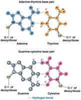
Base Pairing and Hydrogen Bonds
Complementary Base Pairing
Base pairing in DNA is governed by hydrogen bonds:
Adenine (A) pairs with Thymine (T) via two hydrogen bonds
Guanine (G) pairs with Cytosine (C) via three hydrogen bonds
In RNA, uracil (U) replaces thymine and pairs with adenine. This specificity ensures accurate DNA replication and transcription.
DNA vs. RNA: Structural Differences
Strand Structure
DNA is typically double-stranded, forming a stable double helix, while RNA is usually single-stranded but can form short double-stranded regions through intramolecular base pairing. This difference underlies DNA’s role in long-term information storage and RNA’s versatility in cellular functions.
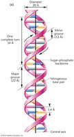
Major and Minor Grooves of DNA
Groove Structure and Protein Interactions
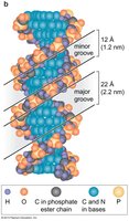
The double helix of DNA forms major and minor grooves due to the geometry of the sugar-phosphate backbones. The major groove (22 Å wide) is more accessible and contains more chemical information for protein binding, while the minor groove (12 Å wide) is narrower and less information-rich. These grooves are critical for DNA-protein interactions, such as those involving transcription factors.
Chargaff’s Rules
Base Composition and Pairing Ratios
Erwin Chargaff’s experiments established that:
The amount of adenine (A) equals thymine (T), and guanine (G) equals cytosine (C) in DNA.
The ratio of purines (A + G) to pyrimidines (C + T) is always approximately 1:1.
Base composition varies between species, but the pairing rules are universal.
These findings provided key evidence for the double helix model and complementary base pairing.
Structural Forms of DNA: B-DNA, A-DNA, and Z-DNA
B-DNA: The Cellular Standard
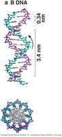
B-DNA is the most common form of DNA under physiological conditions (high humidity). It is a right-handed helix with 10 base pairs per turn, a 34 Å helical pitch, and distinct major and minor grooves.

A-DNA: Dehydrated Form
A-DNA forms under low humidity or partial dehydration. It is also a right-handed helix but is shorter and more compact, with about 11 base pairs per turn, a deeper major groove, and a shallower minor groove. A-DNA is typically observed in DNA–RNA hybrids or in laboratory conditions.
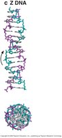
Z-DNA: Left-Handed Helix
Z-DNA is a left-handed helical form that can occur in DNA sequences with alternating purines and pyrimidines. It is less common and may play roles in gene regulation. Other forms (C-DNA, D-DNA, E-DNA, P-DNA) are right-handed and less compact than B-DNA, with D- and E-DNA lacking guanine.
Summary Table: Key Features of DNA Structural Forms
Form | Handedness | Base Pairs/Turn | Major Groove | Minor Groove | Occurrence |
|---|---|---|---|---|---|
B-DNA | Right | 10 | Wide, deep | Narrow, shallow | Physiological conditions |
A-DNA | Right | 11 | Deeper, narrower | Shallower, broader | Dehydrated/lab, DNA–RNA hybrids |
Z-DNA | Left | 12 | Flat | Deep, narrow | Alternating purine-pyrimidine sequences |
DNA Structure: Summary
DNA is composed of four nucleotides: adenine, cytosine, thymine, and guanine.
Base pairing: A = T, G = C; purines = pyrimidines.
Nucleotides are linked by phosphodiester bonds, forming antiparallel strands.
DNA is a right-handed double helix with major and minor grooves.
Hydrogen bonds between bases provide stability and allow strand separation for replication and transcription.
