Back
BackPolysaccharides: Structure, Function, and Biological Roles
Study Guide - Smart Notes
 Tailored notes based on your materials, expanded with key definitions, examples, and context.
Polysaccharides: Structure, Function, and Biological Roles
Introduction to Polysaccharides
Polysaccharides are large, complex carbohydrates composed of long chains of monosaccharide units linked by glycosidic bonds. They play critical roles in energy storage, structural integrity, and cellular communication in living organisms. Polysaccharides can be classified based on their monosaccharide composition and biological function.
Classification of Polysaccharides
Homoglycans and Heteroglycans
Homoglycans (Homopolysaccharides): Polymers consisting of only one type of monosaccharide (e.g., starch, glycogen, cellulose).
Heteroglycans (Heteropolysaccharides): Polymers composed of more than one type of monosaccharide (e.g., glycosaminoglycans, peptidoglycans).
Polysaccharides are synthesized enzymatically without a template, resulting in variable chain lengths. They are classified according to their biological roles, such as storage or structural functions.
Storage Polysaccharides
Starch and Glycogen
Starch (in plants) and glycogen (in animals) are the primary storage polysaccharides, both composed of α-D-glucose units. They differ in their branching patterns and molecular weights.
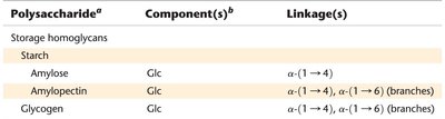
Polysaccharide | Component(s) | Linkage(s) |
|---|---|---|
Starch | Glc | α-(1→4), α-(1→6) (branches) |
Amylose | Glc | α-(1→4) |
Amylopectin | Glc | α-(1→4), α-(1→6) (branches) |
Glycogen | Glc | α-(1→4), α-(1→6) (branches) |
Amylose
Linear polymer of D-glucose with α-(1→4) glycosidic bonds.
Typically contains 100–1000 glucose residues.
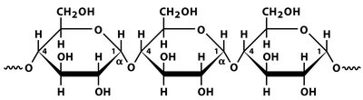
Amylose can adopt a left-handed helical conformation, which is stabilized by hydrogen bonding and hydration.
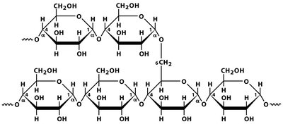
Amylopectin and Glycogen
Amylopectin: Branched polymer with α-(1→4) main chains and α-(1→6) branch points every 24–30 residues.
Glycogen: Similar to amylopectin but with more frequent (every 8–12 residues) and shorter branches, resulting in a higher molecular weight.
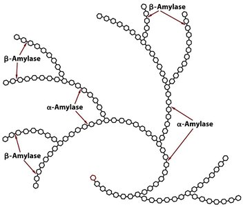
Enzymatic Degradation of Storage Polysaccharides
α-Amylase: Endoglycosidase that hydrolyzes internal α-(1→4) bonds.
β-Amylase: Exoglycosidase that cleaves maltose units from non-reducing ends.
Debranching enzymes: Required to hydrolyze α-(1→6) branch points.
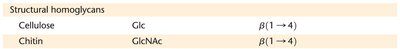
Structural Polysaccharides
Cellulose and Chitin
Structural polysaccharides provide rigidity and strength to cells and tissues. In plants, cellulose is the main structural component, while chitin serves a similar role in fungi and arthropods.
Polysaccharide | Component(s) | Linkage(s) |
|---|---|---|
Cellulose | Glc | β-(1→4) |
Chitin | GlcNAc | β-(1→4) |
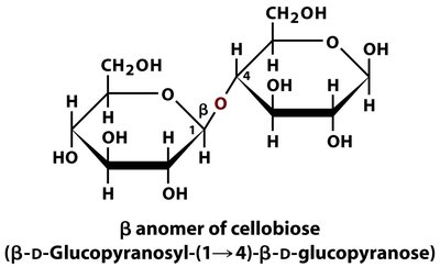
Cellulose
Linear polymer of D-glucose with β-(1→4) glycosidic bonds.
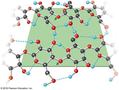
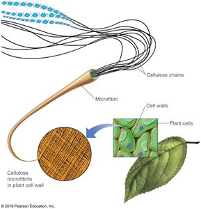
Forms extended chains that associate via hydrogen bonds, resulting in strong microfibrils.
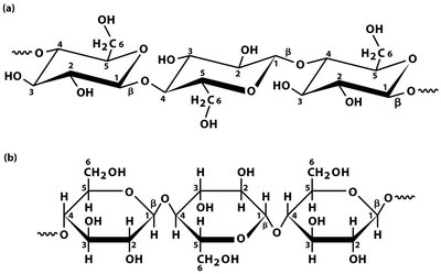
The repeating disaccharide unit of cellulose is cellobiose, linked by β-glycosidic bonds.
Cellulose fibrils are stabilized by extensive intra- and inter-chain hydrogen bonding, contributing to their mechanical strength.
Cellulose microfibrils are the main component of plant cell walls, providing structural support.
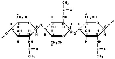
Chitin
Polymer of N-acetyl-β-D-glucosamine (GlcNAc) with β-(1→4) linkages.
Major component of fungal cell walls and arthropod exoskeletons.
Chains are held together by hydrogen bonds, forming strong fibrils.
Glycoconjugates
Proteoglycans, Peptidoglycans, and Glycoproteins
Glycoconjugates are molecules in which polysaccharides are covalently linked to proteins or peptides, playing essential roles in cell signaling, structure, and recognition.
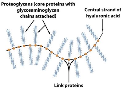
Proteoglycans: Proteins covalently attached to glycosaminoglycans (GAGs), forming major components of the extracellular matrix.
Peptidoglycans: Polysaccharide-peptide complexes found in bacterial cell walls.
Glycoproteins: Proteins with covalently attached oligo- or polysaccharide chains, involved in diverse biological functions.
Glycosaminoglycans (GAGs)
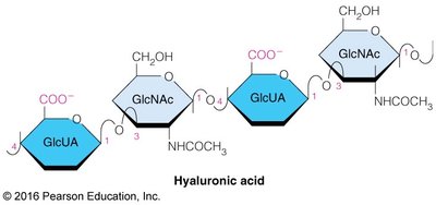
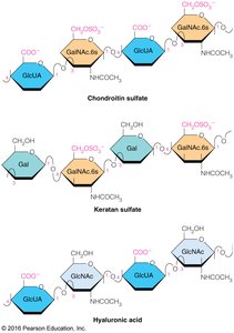
Unbranched heteropolysaccharides composed of repeating disaccharide units (amino sugar + uronic acid).
Often sulfated, resulting in highly negative charges and polyanionic character.
Proteoglycans form complex matrices in tissues such as cartilage, where they provide compressive strength and hydration.
Examples of GAGs include hyaluronic acid, chondroitin sulfate, and keratan sulfate.
Nonstructural Glycosaminoglycans
Hyaluronic acid: Highly soluble, acts as a lubricant in synovial fluid and vitreous humor.
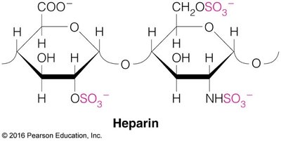
Heparin: A natural anticoagulant that inhibits blood clotting by binding to antithrombin III.
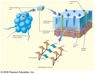
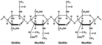
Peptidoglycans
Major component of bacterial cell walls, consisting of alternating N-acetylglucosamine (NAG) and N-acetylmuramic acid (NAM) residues cross-linked by peptides.
Gram-positive bacteria have thick, multilayered peptidoglycan walls; Gram-negative bacteria have a thinner layer covered by an outer membrane.
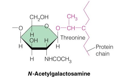
Glycoproteins
Proteins covalently linked to carbohydrate chains, which may be N-linked (to asparagine) or O-linked (to serine or threonine).
Serve as enzymes, hormones, structural proteins, and in cell recognition (e.g., blood group antigens).
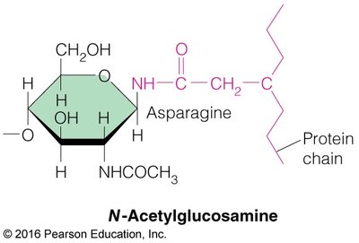
N-linked Glycoproteins
Carbohydrate attached via the amide group of asparagine, typically involving N-acetylglucosamine.
Can have complex, branched structures.
O-linked Glycoproteins
Carbohydrate attached via the hydroxyl group of serine or threonine, typically involving N-acetylgalactosamine.
Important in mucins, blood group antigens, and antifreeze proteins in Antarctic fish.