 Back
Posttranscriptional Regulation in Eukaryotes: Mechanisms and Implications
Study Guide - Smart Notes
 Tailored notes based on your materials, expanded with key definitions, examples, and context.
Posttranscriptional Gene Regulation
Overview of Posttranscriptional Regulation
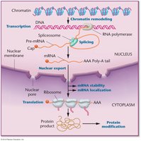
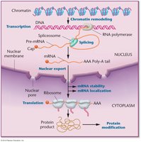
Posttranscriptional regulation encompasses all mechanisms that control gene expression after transcription but before translation. These processes are crucial in eukaryotes, often rivaling transcriptional control in their impact on gene expression.
Key Mechanisms: Alternative splicing, mRNA stability, translation regulation, and RNA silencing.
Importance: Allows cells to fine-tune protein production, respond to environmental cues, and generate protein diversity.
Example: Alternative splicing enables a single gene to produce multiple protein isoforms.
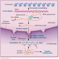
RNA Processing in Eukaryotes
Pre-mRNA Processing
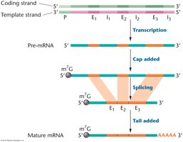
Eukaryotic mRNA is initially transcribed as a precursor (pre-mRNA) that is larger than the mature, translated mRNA. Several modifications are required to produce functional mRNA.
5' Capping: Addition of a 7-methylguanosine cap to the 5' end, protecting mRNA from degradation and aiding translation initiation.
Polyadenylation: Addition of a poly(A) tail to the 3' end, enhancing stability and export from the nucleus.
Splicing: Removal of introns and joining of exons to form mature mRNA.
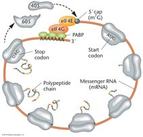
Closed Loop Translation
During translation, mRNA forms a closed loop structure, facilitated by interactions between the cap-binding protein (eIF4E), poly-A binding protein (PABP), and eIF4G. This structure enhances translation efficiency and stability.
Cap and Tail Interaction: Poly-A binding proteins bind to the cap-binding protein, forming the loop.
Translation Initiation: The closed loop promotes ribosome recruitment and translation.
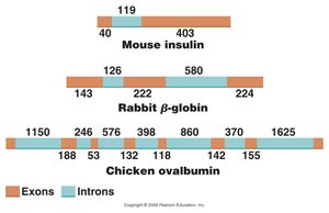
Exons and Introns
Structure of Eukaryotic Genes
Eukaryotic genes are interrupted by introns (non-coding regions) and exons (coding regions). Introns are removed during splicing, while exons are joined to form the mature mRNA.
Exons: Typically 25–300 nucleotides in length; code for protein.
Introns: Range from hundreds to tens of thousands of nucleotides; do not code for protein.
Exon Definition: Exons are defined by their boundaries at the 3' and 5' splice sites.
Gene Size, mRNA Size, and Number of Introns
Gene | Gene Size (kb) | mRNA Size (kb) | Number of Introns |
|---|---|---|---|
Insulin | 1.7 | 0.4 | 2 |
Collagen [pro-α-2(I)] | 38.0 | 5.0 | 50 |
Albumin | 25.0 | 2.1 | 14 |
Phenylalanine hydroxylase | 90.0 | 2.4 | 12 |
Dystrophin | 2000.0 | 17.0 | 50 |
RNA Splicing
Types of Introns and Splicing Mechanisms
Introns are removed from pre-mRNA by different mechanisms, depending on their type.
Group I Introns: The intron RNA acts as a ribozyme, catalyzing its own removal.
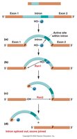
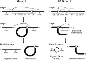
Group II Introns: Splicing occurs via a lariat intermediate, involving two transesterification steps.
Spliceosomal Introns: Most common in nuclear protein-coding genes; require the spliceosome, a complex of snRNPs.
Spliceosome Assembly and Function
The spliceosome recognizes splice sites and catalyzes intron removal.
Recognition: U1 snRNP binds the 5' splice site; U2 binds the branch-point region.
Assembly: U4/U6–U5 complex joins, bringing splice sites together.
Catalysis: U2 and U6 form the catalytic RNA core; U5 aligns exons.
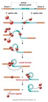
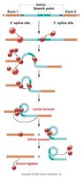
Lariat Formation: Branch-point adenosine's 2'-OH attacks the 5' splice site, forming a lariat.
Exon Joining: Exon 1's 3'-OH attacks the 3' splice site, joining exons and releasing the intron.
Alternative Splicing
Mechanisms and Biological Significance
Alternative splicing allows a single gene to produce multiple mRNA isoforms, increasing protein diversity.
Regulation: Competition among splice sites, influenced by cis-acting elements and trans-acting proteins.
Protein Diversity: At least two-thirds of human protein-coding genes undergo alternative splicing.
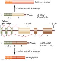
Example: The CT/CGRP gene produces different peptides in thyroid and neuronal cells via alternative splicing.
Regulation of Alternative Splicing
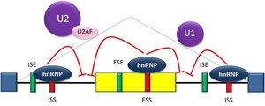
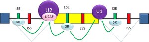
Alternative splicing is regulated by cis-acting RNA sequence elements (enhancers and silencers) and trans-acting RNA-binding proteins (splicing factors).
Silencers and Enhancers: Located in introns (ISS) or exons (ESS).
Splicing Factors: Bind enhancers or silencers, promoting or inhibiting spliceosome assembly.
Thermodynamics and Kinetics: Splicing factors affect both the stability and speed of spliceosome assembly.
Types of Alternative Splicing
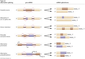
Type of Alternative Splicing | Pre-mRNA | mRNA Isoforms |
|---|---|---|
Cassette exons | Exon can be included or skipped | Isoforms with or without the exon |
Alternative 5' or 3' splice site | Different splice sites used | Isoforms with different exon boundaries |
Intron retention | Intron may be retained | Isoforms with or without the intron |
Mutually exclusive exons | Only one of two exons included | Isoforms with either exon |
Alternative promoters | Different transcription start sites | Isoforms with different 5' ends |
Alternative polyadenylation | Different poly(A) sites used | Isoforms with different 3' ends |
Alternative Splicing in Drosophila Sex Determination
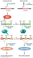
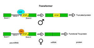
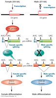
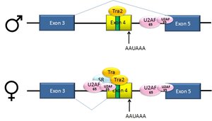
Sex determination in Drosophila is controlled by a cascade of alternative splicing events involving the Sxl, tra, and dsx genes.
Sxl: Splicing repressor; directs splicing of tra pre-mRNA to produce functional Tra protein in females.
Tra: Splicing activator; promotes female-specific dsx splicing.
Dsx: Produces sex-specific transcription factors (DsxF in females, DsxM in males).
Spliceopathies: Genetic Disorders from Splicing Defects
Myotonic Dystrophy
Myotonic dystrophy (DM) is caused by repeat expansions that disrupt splicing regulation.
DM1: CTG repeat expansion in DMPK gene.
DM2: CCTG repeat expansion in CNBP gene.
Pathogenesis: Expanded repeat RNAs interfere with splicing factors, leading to abnormal mRNA processing.
Spinal Muscular Atrophy (SMA)
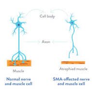
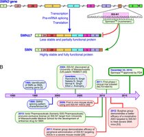
SMA is caused by mutations in the SMN1 gene, leading to loss of motor neurons.
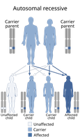
Inheritance: Autosomal recessive; both copies of SMN1 must be mutated.
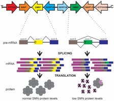
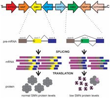
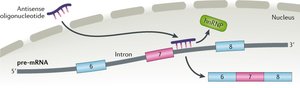
SMN2 Rescue: SMN2 gene produces some functional SMN protein via alternative splicing, but most transcripts skip exon 7.
Spinraza: An antisense oligonucleotide drug that promotes exon 7 inclusion in SMN2, increasing functional SMN protein.
Control of mRNA Stability
mRNA Capping
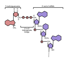
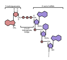
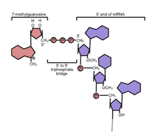
Eukaryotic pre-mRNA receives a 5' cap early during transcription, which protects the mRNA and aids translation.
Structure: 7-methylguanosine (m⁷G) attached via a 5'–5' triphosphate linkage.
Function: Protects from exonucleases, recruits cap-binding proteins, and regulates mRNA decay.
Polyadenylation
Most eukaryotic mRNAs receive a poly(A) tail, which supports stability, translation, and export.
Steps: Recognition of polyadenylation signals, cleavage, and addition of adenosines by poly(A) polymerase.
Function: Poly(A)-binding proteins protect the tail and promote translation.
Alternative Polyadenylation
Cells can select different polyadenylation sites, producing mRNAs with varying regulatory regions.
Short vs. Long Isoforms: Use of proximal or distal poly(A) sites changes the 3' UTR length.
Regulation: Trans-acting factors and cis-elements influence site selection.
mRNA Degradation Pathways
Deadenylation-Dependent Decay: Poly(A) tail shortening leads to mRNA instability and degradation.
Decapping: Removal of the 5' cap exposes mRNA to exonucleases.
Endonucleolytic Cleavage: Creates unprotected ends for exonuclease attack.
Nonsense-Mediated Decay (NMD)
NMD eliminates mRNAs with premature stop codons, preventing production of truncated proteins.
Mechanism: Exon junction complexes (EJCs) mark spliced mRNA; ribosome removes EJCs during translation. If a stop codon is encountered before all EJCs are removed, NMD is triggered.
RNA Silencing and Noncoding RNAs
RNA Interference (RNAi)
Short RNA molecules (siRNA and miRNA) regulate gene expression by repressing translation or triggering mRNA degradation.
siRNA: Derived from double-stranded RNA; guides target RNA cleavage.
miRNA: Derived from RNA hairpins; guides translation repression and mRNA decay.
Long Noncoding RNAs (lncRNA)
lncRNAs (>200nt) are often capped, polyadenylated, and spliced, but lack an open reading frame.
Functions: Modulate alternative splicing, mRNA stability, miRNA sponging, and translational control.
Example: linc-MD1 restores muscle cell differentiation in DMD patients by sponging miRNAs.
Translational and Posttranslational Regulation
Cytoplasmic Polyadenylation and mRNA Localization
Cytoplasmic Polyadenylation: Regulates translation initiation and mRNA localization.
mRNA Localization: Zip code elements in the 3' UTR direct mRNA to specific cellular locations, where translation is activated.
Posttranslational Modifications
Phosphorylation: Addition of phosphate groups by kinases; removal by phosphatases. Alters protein function and activity.
Ubiquitination: Targets proteins for degradation by the proteasome.
Example: p53 protein is regulated by ubiquitin-mediated degradation; DNA damage inhibits degradation, allowing p53 to activate DNA repair genes.
Summary Table: Key Concepts in Posttranscriptional Regulation
Process | Key Features | Biological Significance |
|---|---|---|
Alternative Splicing | Multiple mRNA isoforms from one gene | Protein diversity, cell differentiation |
mRNA Stability | Regulated by capping, polyadenylation, RBPs | Controls protein abundance |
RNA Silencing | siRNA, miRNA, lncRNA | Gene expression regulation, antiviral defense |
Translational Control | Closed loop translation, cytoplasmic polyadenylation | Timing and localization of protein synthesis |
Posttranslational Modification | Phosphorylation, ubiquitination | Protein activity, degradation |
