 Back
Posttranscriptional Regulation in Eukaryotes: Mechanisms and Implications
Study Guide - Smart Notes
 Tailored notes based on your materials, expanded with key definitions, examples, and context.
Posttranscriptional Gene Regulation
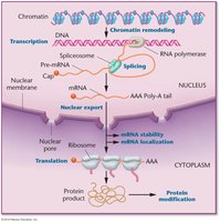
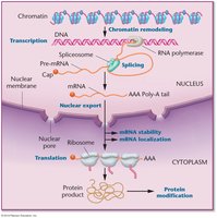
Overview of Posttranscriptional Regulation
Posttranscriptional regulation refers to the control of gene expression after transcription has occurred, encompassing all processes that modify or influence the fate of RNA transcripts. In eukaryotes, these mechanisms are essential for generating protein diversity, regulating gene expression, and responding to cellular signals.
Key Mechanisms: Alternative splicing, mRNA stability, translation control, and RNA silencing.
Significance: These processes allow a single gene to produce multiple protein isoforms and enable rapid adaptation to environmental or developmental cues.
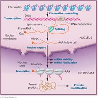
RNA Processing in Eukaryotes
Pre-mRNA Processing Steps
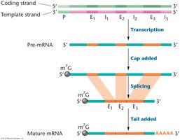
Eukaryotic mRNA is initially synthesized as a precursor (pre-mRNA) that undergoes several modifications before becoming a mature, translatable mRNA.
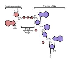
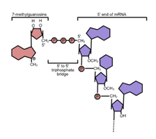
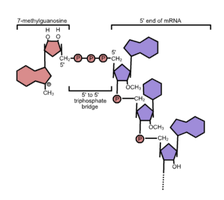
5' Capping: Addition of a 7-methylguanosine cap to the 5' end, protecting the RNA and facilitating translation initiation.
Splicing: Removal of non-coding introns and joining of coding exons.
3' Polyadenylation: Addition of a poly(A) tail to the 3' end, enhancing stability and export.
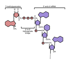
5' Cap Structure
The 5' cap structure is a unique feature of eukaryotic mRNAs, consisting of a 7-methylguanosine linked via a 5'-5' triphosphate bridge to the first nucleotide of the RNA.
Function: Protects mRNA from exonuclease degradation and is recognized by cap-binding proteins during translation initiation.
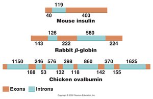
Exons and Introns
Genes in eukaryotes are typically interrupted by introns (non-coding regions) interspersed among exons (coding regions). The length and number of introns and exons can vary widely among genes and species.
Exons: Typically 25–300 nucleotides in humans; encode protein sequences.
Introns: Range from hundreds to tens of thousands of nucleotides; removed during splicing.
RNA Splicing Mechanisms
Types of Introns
Introns are classified based on their splicing mechanisms:
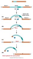
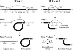
Group I Introns: Self-splicing ribozymes; use a free guanosine as a cofactor.
Group II Introns: Self-splicing; form a lariat intermediate via a branch-point adenosine.
Spliceosomal Introns: Require the spliceosome, a complex of snRNPs and proteins, for removal; most common in nuclear genes.
Spliceosome-Mediated Splicing
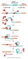
The spliceosome recognizes conserved sequences at the 5' (GU) and 3' (AG) splice sites and the branch-point adenosine. Splicing occurs via two transesterification reactions, resulting in exon ligation and intron removal as a lariat.
snRNPs: U1, U2, U4, U5, and U6 are essential for splice site recognition and catalysis.
Splicing Steps: Assembly, activation, lariat formation, exon ligation, and disassembly.
Alternative Splicing
Mechanisms and Biological Significance
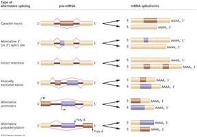
Alternative splicing allows a single gene to produce multiple mRNA and protein isoforms by varying the combination of exons included in the mature transcript. This process greatly expands proteomic diversity without increasing gene number.
Regulation: Controlled by cis-acting elements (enhancers/silencers) and trans-acting splicing factors (e.g., SR proteins, hnRNPs).
Prevalence: Over two-thirds of human protein-coding genes undergo alternative splicing.
Example: Calcitonin/CGRP Gene
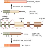
The calcitonin/CGRP gene produces two distinct peptides via alternative splicing and polyadenylation in different tissues.
Thyroid cells: Include exon 4, producing calcitonin.
Neuronal cells: Include exons 5 and 6, producing CGRP.
Regulation of Alternative Splicing
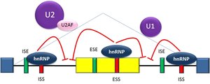
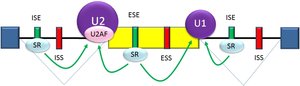
Alternative splicing is regulated by the interplay of cis-acting RNA elements and trans-acting proteins. Enhancers and silencers can be located in exons or introns, and their binding by specific proteins determines splice site usage.
Enhancers: Exonic (ESE) or intronic (ISE) sequences that promote splicing.
Silencers: Exonic (ESS) or intronic (ISS) sequences that repress splicing.
Splicing Factors: SR proteins (activators) and hnRNPs (repressors) modulate spliceosome assembly and activity.
Alternative Splicing in Drosophila Sex Determination
Sex Determination Pathway
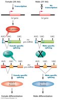
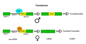
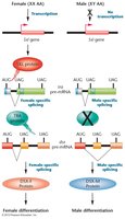
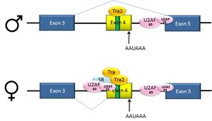
In Drosophila, sex is determined by the ratio of X chromosomes to autosomes, interpreted by a cascade of alternative splicing events involving the Sex lethal (Sxl), transformer (tra), and doublesex (dsx) genes.
Female (XX): Sxl protein is produced, leading to functional Tra protein and female-specific DsxF isoform.
Male (XY): No Sxl protein, default splicing produces truncated Tra and male-specific DsxM isoform.
Spliceopathies: Genetic Disorders of Splicing
Myotonic Dystrophy
Myotonic dystrophy (DM) is a spliceopathy caused by repeat expansions in the DMPK (DM1) or CNBP (DM2) genes, leading to disrupted splicing regulation and multisystemic symptoms.
DM1: CTG repeat expansion in DMPK gene.
DM2: CCTG repeat expansion in CNBP gene.
Spinal Muscular Atrophy (SMA)
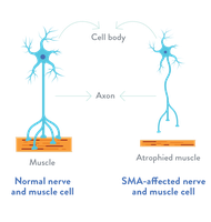
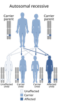
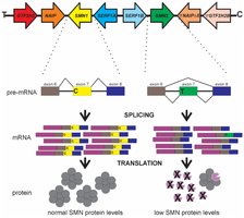
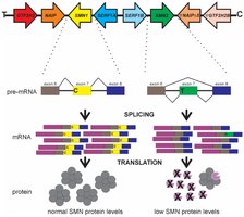
SMA is an autosomal recessive neuromuscular disorder caused by loss of function in both copies of the SMN1 gene. The nearly identical SMN2 gene can partially compensate, but a single nucleotide change leads to exon 7 skipping in most transcripts, producing unstable protein.
Inheritance: Autosomal recessive; both parents must carry the mutation.
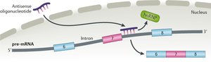
Therapy: Spinraza (nusinersen) is an antisense oligonucleotide that promotes exon 7 inclusion in SMN2 transcripts, restoring functional SMN protein.
Control of mRNA Stability
mRNA Capping and Polyadenylation
mRNA stability is influenced by the presence of a 5' cap and a 3' poly(A) tail. The cap protects against 5'→3' exonucleases, while the poly(A) tail and associated binding proteins protect against 3'→5' degradation.
Decapping: Removal of the cap exposes the mRNA to degradation by XRN1.
Deadenylation: Shortening of the poly(A) tail precedes mRNA decay.
Alternative Polyadenylation
Many genes have multiple polyadenylation sites, allowing the production of mRNA isoforms with different 3' UTRs. This can affect mRNA stability, localization, and translation by including or excluding regulatory elements.
Regulation: Controlled by processing factors such as CstF64 and sequence elements in the pre-mRNA.
mRNA Surveillance and Decay Pathways
Nonsense-Mediated Decay (NMD)
NMD is a quality control mechanism that degrades mRNAs containing premature termination codons (PTCs), preventing the production of truncated, potentially harmful proteins.
Mechanism: Exon junction complexes (EJCs) mark splice sites; if translation terminates upstream of an EJC, NMD is triggered.
RNA Silencing and Noncoding RNAs
RNA Interference (RNAi)
Small noncoding RNAs, including siRNAs and miRNAs, regulate gene expression posttranscriptionally by guiding the RNA-induced silencing complex (RISC) to target mRNAs for degradation or translational repression.
siRNAs: Derived from long double-stranded RNA; direct cleavage of perfectly complementary targets.
miRNAs: Derived from hairpin precursors; typically repress translation or promote decay via partial complementarity.
Long Noncoding RNAs (lncRNAs)
lncRNAs are transcripts longer than 200 nucleotides that do not encode proteins but regulate gene expression through diverse mechanisms, including chromatin remodeling, splicing modulation, mRNA stability, and miRNA sponging.
Classes: Antisense, intronic, bidirectional, and intergenic lncRNAs.
Functions: Guide chromatin modifiers, act as decoys, scaffolds, or guides, and regulate mRNA processing and translation.
Translational and Posttranslational Regulation
Cytoplasmic Polyadenylation and mRNA Localization
Some mRNAs are stored in a translationally repressed state and activated by cytoplasmic polyadenylation in response to specific signals. Localization elements (zip codes) in the 3' UTR direct mRNAs to specific cellular regions, where translation is locally activated.
Posttranslational Modifications
Protein activity and stability are further regulated after translation by modifications such as phosphorylation and ubiquitination.
Phosphorylation: Addition of phosphate groups by kinases; removal by phosphatases. Alters protein conformation and function.
Ubiquitination: Covalent attachment of ubiquitin targets proteins for degradation by the proteasome.
Regulation of p53 Protein
The tumor suppressor p53 is regulated by ubiquitin-mediated degradation. Under normal conditions, Mdm2 ubiquitinates p53, targeting it for proteasomal degradation. Upon DNA damage, Mdm2 is phosphorylated and loses affinity for p53, allowing p53 accumulation and activation of cell cycle arrest and DNA repair genes.
