 Back
BackChapter 20
Study Guide - Smart Notes
 Tailored notes based on your materials, expanded with key definitions, examples, and context.
Chapter 20: Recombinant DNA Technology
Introduction
Recombinant DNA technology has revolutionized genetics by enabling the manipulation, amplification, and analysis of DNA and RNA. This chapter focuses on the polymerase chain reaction (PCR), molecular techniques for analyzing nucleic acids, and DNA sequencing methods, all of which are foundational for modern genetic research and biotechnology.
The Polymerase Chain Reaction (PCR)
Discovery and Historical Context
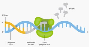
The development of PCR was made possible by the discovery of heat-stable DNA polymerase from the thermophilic bacterium Thermus aquaticus, found in Yellowstone National Park. This enzyme, known as Taq polymerase, can withstand the high temperatures required for DNA denaturation during PCR cycles.

Principle and Steps of PCR
PCR is a technique used to amplify specific DNA sequences in vitro, allowing for the generation of millions of copies from a small initial sample. The process involves three main steps, repeated in cycles:
Denaturation: Double-stranded DNA is separated into single strands by heating (typically 94–98°C).
Primer Annealing: Short DNA primers bind to complementary sequences on the single-stranded DNA at a lower temperature (50–65°C).
Extension: Taq polymerase synthesizes new DNA strands by adding nucleotides (dNTPs) to the primers at 72°C.
Each cycle doubles the amount of target DNA, resulting in exponential amplification.
Applications of PCR
Detection of genetic mutations
Cloning of DNA fragments
Forensic analysis and paternity testing
Diagnosis of infectious diseases
Gene expression analysis (with modifications such as RT-PCR)
Example: PCR is used to detect the presence of pathogenic DNA in clinical samples, such as identifying viral infections.
Reverse Transcription PCR (RT-PCR) and Quantitative PCR (qPCR)
RT-PCR: Studying Gene Expression
RT-PCR is used to study gene expression by converting messenger RNA (mRNA) into complementary DNA (cDNA) using the enzyme reverse transcriptase. The cDNA can then be amplified by PCR, allowing researchers to detect and analyze gene expression patterns.
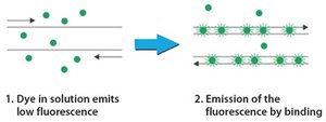
Quantitative Real-Time PCR (qPCR)
qPCR enables the quantification of DNA or cDNA during the PCR process in real time, often using fluorescent dyes such as SYBR Green. The increase in fluorescence correlates with the amount of DNA amplified, allowing for precise measurement of gene expression levels.
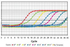
Key Point: qPCR is highly sensitive and can detect small differences in gene expression between samples.
Key Point: The threshold cycle (Ct) value is inversely proportional to the initial amount of target nucleic acid.
Example: qPCR is used in medical diagnostics to quantify viral load in patient samples.
Molecular Techniques for Analyzing DNA and RNA
Blotting Techniques
Blotting techniques are used to detect specific nucleic acids or proteins separated by electrophoresis:
Southern blot: Detects DNA
Northern blot: Detects RNA
Western blot: Detects proteins
All methods involve transferring molecules to a membrane and probing with labeled complementary sequences or antibodies.
RNA In Situ Hybridization
This technique allows for the visualization of gene expression within tissues. A labeled RNA probe binds to the target mRNA in cells, and detection is achieved through antibody binding and a colorimetric reaction, resulting in stained cells where the gene is expressed.
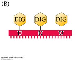
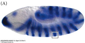
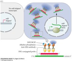
Key Point: In situ hybridization provides spatial information about gene expression within tissues or embryos.
Immunohistochemistry (IHC)
IHC is used to detect specific proteins in tissue sections using antibodies. A primary antibody binds to the protein of interest, and a secondary antibody (conjugated to an enzyme or fluorescent molecule) enables visualization.
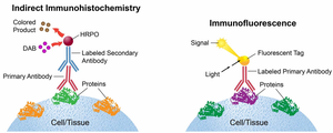
Key Point: IHC can reveal the localization and abundance of proteins in cells and tissues.
Key Point: Fluorescent tags allow for multiplexing and co-localization studies.
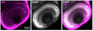
Example: Immunohistochemistry in Zebrafish
Whole-mount immunostaining can be used to visualize the expression of proteins such as glutamine synthetase in zebrafish retina, with DNA counterstained using DAPI.
RNA Sequencing (RNA-Seq)
Principle and Applications
RNA-Seq is a high-throughput sequencing technique used to analyze the transcriptome—the complete set of RNA transcripts in a cell or tissue at a given time. It allows for the comparison of gene expression across different tissues, developmental stages, or experimental conditions.
Key Point: RNA-Seq can detect both coding and non-coding RNAs, including microRNAs.
Key Point: Requires significant computational analysis to process and interpret large datasets.
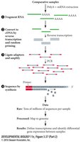
Example: RNA-Seq can identify genes that are differentially expressed in cancerous versus normal tissues.
DNA Sequencing
Sanger Sequencing
Sanger sequencing, or dideoxynucleotide chain-termination sequencing, uses modified nucleotides (dideoxynucleotides) that terminate DNA synthesis. By incorporating these chain-terminators, the sequence of DNA can be determined based on the lengths of terminated fragments.
Key Point: Sanger sequencing is accurate but limited in throughput compared to newer methods.
Next-Generation and Third-Generation Sequencing
Modern sequencing technologies allow for massively parallel sequencing of millions of DNA fragments, greatly reducing the cost and time required to sequence entire genomes. Third-generation sequencing can read long stretches of DNA from single molecules, further advancing genomics research.
Key Point: The cost of sequencing a human genome has dropped dramatically, making personalized genomics feasible.
Summary Table: Comparison of Gene Expression Analysis Techniques
Technique | Analyte | Quantitative? | Spatial Information? | Throughput |
|---|---|---|---|---|
qPCR | mRNA (via cDNA) | Yes | No | Low |
RNA in situ hybridization | mRNA | No | Yes | Low |
RNA-Seq | All RNAs | Yes | No | High |
Western blot | Protein | Semi-quantitative | No | Low |
Immunohistochemistry | Protein | No | Yes | Low |
Key Equations
PCR Amplification: The number of DNA copies after n cycles is given by: where is the initial number of DNA molecules and is the number of cycles.
Conclusion
Recombinant DNA technology and molecular analysis techniques are essential tools in genetics, enabling the amplification, detection, and sequencing of nucleic acids. These methods have broad applications in research, medicine, and biotechnology, and continue to evolve with advances in technology.
