 Back
BackRecombinant DNA Technology: Tools, Methods, and Applications
Study Guide - Smart Notes
 Tailored notes based on your materials, expanded with key definitions, examples, and context.
Recombinant DNA Technology
Introduction to Recombinant DNA
Recombinant DNA technology, also known as gene splicing, involves creating DNA molecules by joining together genetic material from different sources. This technology allows scientists to isolate, study, and manipulate specific DNA sequences, enabling advances in genetics, biotechnology, and medicine.
Clones: Recovered copies of recombinant DNA molecules used to study DNA structure and function.
Applications: Gene cloning, genetic engineering, and the production of genetically modified organisms (GMOs).
Tools of Recombinant DNA Technology
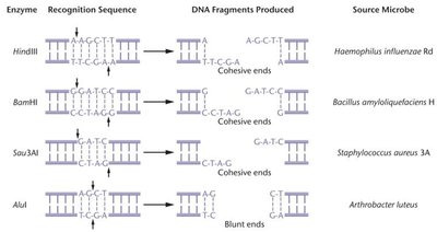
Restriction Enzymes
Restriction enzymes are DNA-cutting enzymes produced by bacteria as a defense mechanism against bacteriophage infection. They recognize specific DNA sequences (restriction sites) and cleave both DNA strands, generating restriction fragments.
Recognition Sequence: Specific palindromic DNA sequences where enzymes bind and cut.
Types of Ends:
Sticky (cohesive) ends: Overhanging single-stranded ends that can anneal with complementary sequences.
Blunt ends: Double-stranded ends with no overhangs.
Example: EcoRI produces sticky ends, while AluI produces blunt ends.
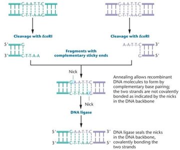
DNA Ligase
DNA ligase is an enzyme that covalently joins DNA fragments by sealing the phosphodiester backbone, forming intact recombinant DNA molecules.
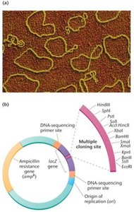
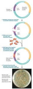
Cloning Vectors
Definition and Features
Cloning vectors are DNA molecules that accept foreign DNA fragments and replicate them in host cells. Essential features include:
Ability to replicate independently of the host chromosome
Multiple restriction enzyme sites (multiple cloning site, MCS)
Selectable marker genes (e.g., antibiotic resistance)
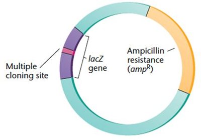
Bacterial Plasmid Vectors
Plasmids are small, circular DNA molecules used as vectors in bacterial cloning. They are engineered to contain MCS, selectable markers, and origins of replication.
Other Types of Cloning Vectors
Phage vectors: Modified bacteriophage genomes, can carry larger DNA fragments (up to 45 kb).
Bacterial Artificial Chromosomes (BACs): Large plasmids for cloning 100–300 kb DNA fragments.
Yeast Artificial Chromosomes (YACs): Vectors for cloning up to 1000 kb, containing telomeres, centromeres, and origins of replication.
Expression Vectors
Expression vectors are designed to ensure transcription and translation of cloned genes, enabling protein production in host cells. They are available for both prokaryotic and eukaryotic systems.
Transformation and Selection
Transformation Methods
Transformation is the process of introducing recombinant plasmids into bacterial cells. Common methods include:
Calcium chloride and heat shock
Electroporation (high-intensity electric pulse)
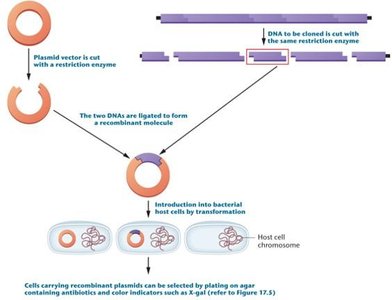
Blue-White Screening
Blue-white screening distinguishes recombinant from nonrecombinant plasmids using the lacZ gene and X-gal substrate:
Functional lacZ (no insert): Blue colonies (can metabolize X-gal)
Disrupted lacZ (with insert): White colonies (cannot metabolize X-gal)
Genomic and cDNA Libraries
Genomic Libraries
A genomic library contains overlapping DNA fragments representing the entire genome of an organism. Constructed by cutting genomic DNA with restriction enzymes and cloning into vectors.
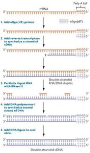
cDNA Libraries
A cDNA library is made from mRNA isolated from cells, representing only expressed genes. Reverse transcriptase synthesizes cDNA, which is then cloned into vectors.
Library Screening
Library screening uses labeled DNA or RNA probes to identify and isolate specific genes of interest from a library.
Polymerase Chain Reaction (PCR)
PCR Principles and Requirements
PCR is a rapid, in vitro method for amplifying specific DNA sequences without the need for host cells. Requirements include:
Double-stranded target DNA
DNA polymerase (e.g., Taq polymerase)
Primers (short, single-stranded DNA sequences)
Deoxyribonucleoside triphosphates (dNTPs)
Mg2+ as a cofactor
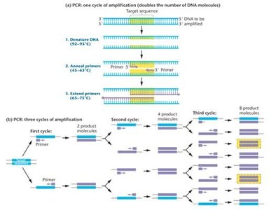
PCR Steps
Denaturation: DNA strands are separated by heating.
Annealing: Primers bind to complementary sequences.
Extension: DNA polymerase synthesizes new DNA strands.
Each cycle doubles the amount of target DNA, resulting in exponential amplification.
Limitations and Applications
Requires prior knowledge of target sequence for primer design.
Highly sensitive to contamination.
Cannot amplify very long DNA segments.
Applications: Genetic testing, forensics, molecular diagnostics.
RT-PCR and qPCR
RT-PCR: Reverse transcription PCR for studying gene expression by converting mRNA to cDNA.
qPCR: Quantitative real-time PCR for measuring DNA amplification in real time.
DNA Analysis Techniques
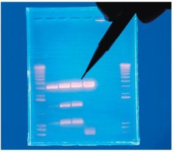
Agarose Gel Electrophoresis
Agarose gel electrophoresis separates DNA fragments by size. Smaller fragments migrate farther through the gel. DNA is visualized using stains like ethidium bromide under UV light.
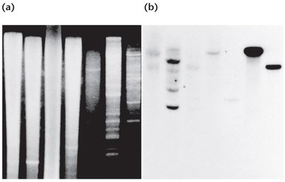
Southern, Northern, and Western Blotting
Southern blot: Detects specific DNA sequences by hybridization with labeled probes after gel electrophoresis and transfer to a membrane.
Northern blot: Analyzes RNA to study gene expression patterns.
Western blot: Detects specific proteins using antibodies.
Fluorescent in Situ Hybridization (FISH)
FISH uses fluorescently labeled probes to hybridize directly to chromosomes or RNA in cells or tissues, allowing visualization of gene expression or chromosomal location.
DNA Sequencing
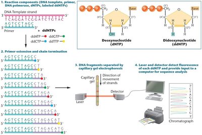
Sanger Sequencing (Dideoxynucleotide Chain-Termination)
The Sanger method uses dideoxynucleotides (ddNTPs) to terminate DNA synthesis at specific bases, generating fragments that can be separated and read to determine the DNA sequence.
Genome Editing: CRISPR-Cas9
CRISPR-Cas9 Mechanism
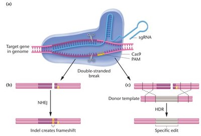
CRISPR-Cas9 is a powerful genome editing tool derived from bacterial immune systems. The Cas9 nuclease, guided by a single guide RNA (sgRNA), introduces double-stranded breaks at specific genomic locations adjacent to a protospacer adjacent motif (PAM) sequence (5'-NGG-3').
Repair mechanisms:
Nonhomologous end-joining (NHEJ): Can introduce insertions or deletions (indels), disrupting gene function.
Homology-directed repair (HDR): Uses a donor template to introduce specific edits.
Limitations of CRISPR-Cas9
Potential for off-target effects due to imperfect sgRNA binding.
Ongoing improvements include engineered Cas9 variants and optimized sgRNA design.
