 Back
BackRegulation of Gene Expression in Bacteria and Bacteriophage
Study Guide - Smart Notes
 Tailored notes based on your materials, expanded with key definitions, examples, and context.
Regulation of Gene Expression in Bacteria and Bacteriophage
Transcriptional Control and DNA–Protein Interactions
Gene expression in bacteria is primarily regulated at the transcriptional level, involving interactions between DNA-binding proteins and regulatory DNA sequences. Some genes are constitutively transcribed for routine cellular functions, while others are regulated in response to environmental changes.
Constitutive transcription: Continuous expression of genes required for basic cellular processes.
Regulated transcription: Genes are transcribed only under specific conditions, allowing adaptation to environmental changes.
Regulation mechanisms: Control of transcription initiation and the amount of transcription.
Mechanisms for Regulating Bacterial Gene Expression
Bacterial gene expression is regulated at both transcriptional and post-transcriptional levels. The main mechanisms are summarized below:
Regulation Type | Mechanism | Example |
|---|---|---|
Transcriptional Regulation | Inducible transcription | lac operon |
Transcriptional Regulation | Repressible transcription | trp operon |
Transcriptional Regulation | Attenuation | trp operon, riboswitches |
Posttranscriptional Regulation | mRNA destruction | riboswitches |
Posttranscriptional Regulation | Translation blockage | antisense RNA, riboswitches |
Negative and Positive Control of Transcription
Transcriptional regulation can be negative or positive, depending on the role of regulatory proteins:
Negative control: A repressor protein binds to a regulatory DNA sequence (operator) and prevents transcription.
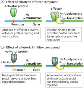
Positive control: An activator protein binds to a regulatory DNA sequence (activator binding site) and facilitates transcription initiation.
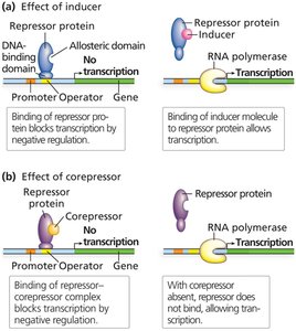
Repressor Proteins and Allostery
Repressor proteins exert negative control and typically have two active sites:
DNA-binding domain: Binds to operator or other regulatory DNA sequences.
Allosteric domain: Binds small molecules (inducers or corepressors), causing conformational changes that affect DNA binding (allostery).
Allostery can either inactivate or activate the DNA-binding domain, depending on the type of effector molecule.
Activator Proteins and Positive Control
Activator proteins facilitate transcription by binding to activator binding sites and helping RNA polymerase bind to the promoter. Their activity is also regulated allosterically by effector or inhibitor compounds.
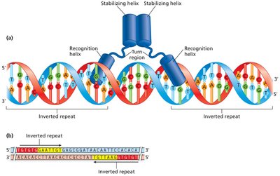
Structural Features of DNA-Binding Proteins
DNA-binding proteins often contain motifs such as the helix-turn-helix (HTH), which allows specific interaction with DNA sequences, often at inverted or direct repeats. These proteins may function as homodimers or heterodimers.
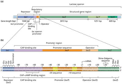
The lac Operon: An Inducible Operon System
Overview and Structure
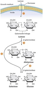
The lac operon in E. coli is a classic example of an inducible operon system, responsible for the metabolism of lactose. It consists of a regulatory region and three structural genes:
lacZ: Encodes β-galactosidase (breaks down lactose).
lacY: Encodes permease (transports lactose into the cell).
lacA: Encodes transacetylase (protects against harmful by-products).
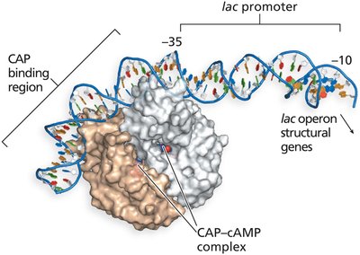
The regulatory region includes the promoter (binds RNA polymerase), operator (binds lac repressor), and CAP binding site (binds CAP–cAMP complex).
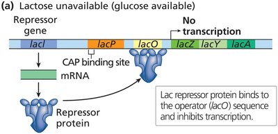
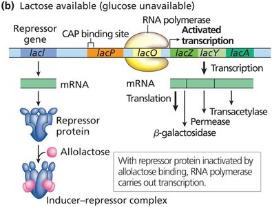
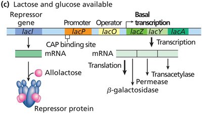
Regulation of the lac Operon
The lac operon is subject to both negative and positive control:
Negative control: In the absence of lactose, the lac repressor binds to the operator, blocking transcription.
Induction: When lactose is present and glucose is absent, allolactose (an isomer of lactose) binds to the repressor, inactivating it and allowing transcription.
Positive control: The CAP–cAMP complex binds to the CAP site, enhancing transcription when glucose is absent.
Mutational Analysis of the lac Operon
Mutational studies have identified the roles of various genes and regulatory sequences in the lac operon. Key mutations include:
Gene/Sequence | Product/Function | Important Mutants |
|---|---|---|
lacI | Repressor protein; binds operator and allolactose | I−: Cannot bind operator (constitutive expression); IS: Super-repressor, cannot bind inducer (no expression) |
lacZ | β-galactosidase | Z−: No functional enzyme |
lacY | Permease | Y−: No functional permease |
lacA | Transacetylase | A−: No transacetylase |
lacO | Operator | OC: Cannot bind repressor (constitutive expression) |
lacP | Promoter | P−: Cannot bind RNA polymerase (no expression) |
The trp Operon: A Repressible and Attenuated System
Overview and Structure
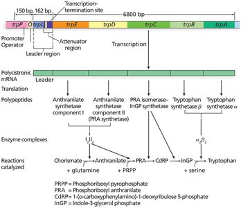
The trp operon in E. coli is a repressible operon responsible for the biosynthesis of tryptophan. It contains five structural genes (trpE, trpD, trpC, trpB, trpA) and a regulatory region with a promoter, operator, and leader/attenuator region.
Regulation by Repression and Attenuation
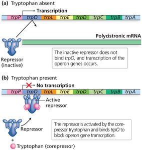
Repression: When tryptophan is abundant, it acts as a corepressor, binding to the trp repressor protein, which then binds the operator and blocks transcription.
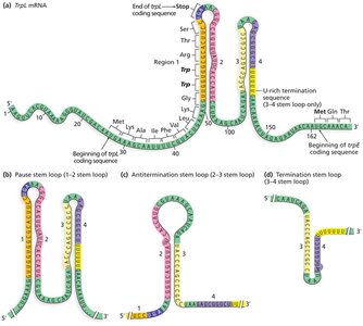
Attenuation: A secondary regulatory mechanism involving the formation of alternative stem-loop structures in the leader mRNA, which can terminate or allow transcription based on tryptophan availability.
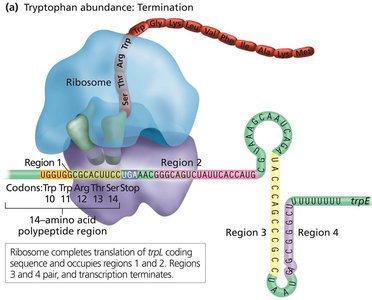
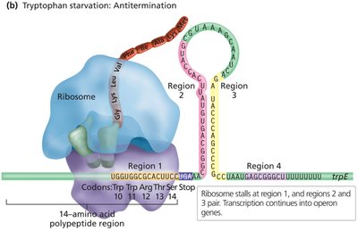
Mechanism of Attenuation
The leader region contains four repeat sequences that can form different stem-loop structures:
3–4 stem loop: Terminates transcription (when tryptophan is abundant).
2–3 stem loop: Antitermination structure, allows transcription (when tryptophan is scarce).

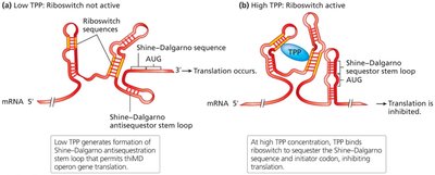
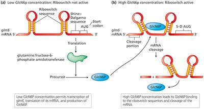
Riboswitches: Regulation Beyond Proteins
Riboswitches in Bacterial Gene Regulation
Riboswitches are regulatory segments of mRNA that bind small molecules, leading to changes in mRNA structure and affecting transcription, translation, or mRNA stability.
Transcriptional regulation: Binding of a metabolite (e.g., TPP) to the riboswitch can cause formation of a termination stem-loop, halting transcription.
Translational regulation: Riboswitches can sequester the Shine-Dalgarno sequence, preventing ribosome binding and translation initiation.
mRNA stability: Some riboswitches induce mRNA cleavage in response to metabolite binding, reducing mRNA stability and translation.
Summary Table: Key Regulatory Mechanisms in Bacteria
Mechanism | Type | Example | Key Features |
|---|---|---|---|
Inducible Operon | Negative/Positive | lac operon | Induced by substrate (allolactose); repressor inactivated; CAP–cAMP positive control |
Repressible Operon | Negative | trp operon | Repressed by end product (tryptophan); corepressor activates repressor |
Attenuation | Transcriptional | trp operon | Leader mRNA forms stem-loops; regulates transcription based on amino acid availability |
Riboswitch | Transcriptional/Translational/Post-transcriptional | thi, glmS operons | mRNA binds small molecule; alters structure to regulate gene expression |
