 Back
BackStructure and Function of Cell Membranes: Genetics Study Guide
Study Guide - Smart Notes
 Tailored notes based on your materials, expanded with key definitions, examples, and context.
Cell Membrane Structure and Function
Overview of Cell Membranes
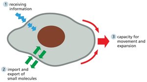
Cell membranes are fundamental to all living cells, acting as selective barriers that separate cellular components from the external environment. Their structure and function are essential for maintaining cellular integrity, facilitating communication, and regulating the transport of molecules.
Selective Barrier: Prevents uncontrolled interaction between cell contents and surroundings.
Exchange of Nutrients and Waste: Specialized channels and transporters allow import and export of molecules.
Environmental Sensing: Membrane receptors enable cells to receive information from their environment.
Flexibility: Membranes allow cells to grow, change shape, and move.
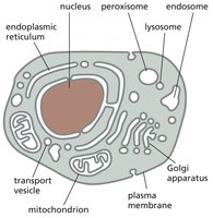
Internal Membranes and Cellular Compartments
Eukaryotic cells contain internal membranes that create distinct compartments, each with specialized functions. In contrast, bacteria and archaea only possess a plasma membrane.
Organelles: Internal membranes enclose organelles such as the nucleus, mitochondria, endoplasmic reticulum, Golgi apparatus, lysosomes, and peroxisomes.
Compartmentalization: Enables separation of metabolic processes and efficient cellular function.
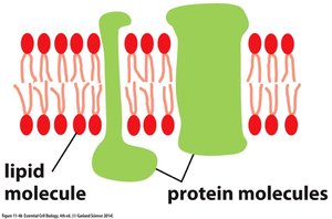
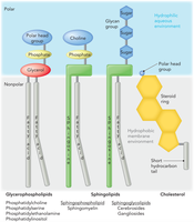
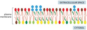
Composition and Organization of Cell Membranes
Lipid Bilayer and Membrane Proteins
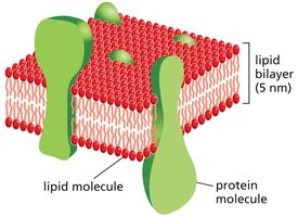
All cell membranes share a common structure: a lipid bilayer with proteins embedded or associated.
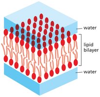
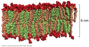
Lipid Bilayer: Composed primarily of phospholipids, forming a double layer about 5 nm thick.
Membrane Proteins: Inserted into or attached to the bilayer, responsible for most membrane functions.
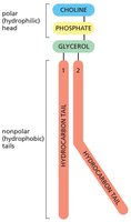
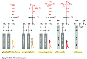
Phospholipids: Structure and Properties
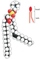
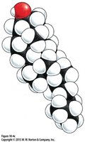
Phospholipids are amphipathic molecules, meaning they contain both hydrophilic (water-loving) and hydrophobic (water-fearing) regions.
Hydrophilic Head: Composed of choline, phosphate, and glycerol.
Hydrophobic Tails: Two hydrocarbon chains.
Phosphatidylcholine: The most common phospholipid in cell membranes.
Major Phospholipids in Mammalian Membranes
Mammalian plasma membranes contain four major phospholipids, each with distinct head groups and fatty acid tails.
Phosphatidylethanolamine
Phosphatidylserine
Phosphatidylcholine
Sphingomyelin
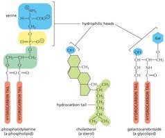
Other Membrane Lipids
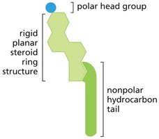
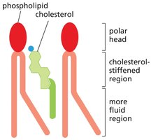
Membranes also contain glycolipids and sterols (such as cholesterol), which contribute to membrane structure and function.
Glycolipids: Lipids with carbohydrate groups, important for cell recognition.
Cholesterol: Modulates membrane fluidity and stability.
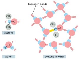
Amphipathic Nature and Bilayer Formation
Hydrophilic vs. Hydrophobic Interactions
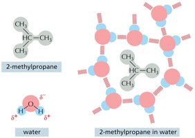
Amphipathic molecules are subject to two conflicting forces: hydrophilic regions interact with water, while hydrophobic regions avoid water.
Hydrophilic Molecules: Dissolve in water via electrostatic attractions and hydrogen bonds.
Hydrophobic Molecules: Cluster together to minimize energy cost, forming cage-like structures in water.
Fat Molecules vs. Phospholipids
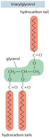
Fat molecules (triacylglycerols) are purely hydrophobic and form large droplets in water, while phospholipids are amphipathic and form bilayers.
Triacylglycerol: Main component of animal fats and plant oils, with three hydrocarbon tails.
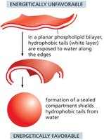
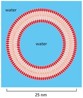
Bilayer Formation and Stability
Amphipathic phospholipids spontaneously form bilayers in water, with hydrophilic heads facing outward and hydrophobic tails shielded inside.
Energetically Favorable: Bilayer formation avoids exposure of hydrophobic tails to water.
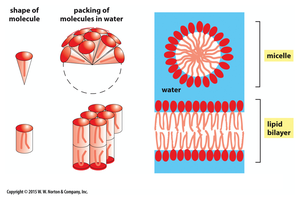
Packing Arrangements: Micelles vs. Bilayers
The shape of amphipathic molecules determines whether they form micelles (cone-shaped) or bilayers (cylinder-shaped) in water.
Micelles: Spherical structures formed by cone-shaped lipids.
Bilayers: Sheet-like structures formed by cylinder-shaped lipids.
Self-Sealing Properties of Bilayers
Phospholipid bilayers are self-sealing, forming closed compartments to avoid free edges and exposure of hydrophobic tails.
Stable Structure: Bilayers bend and seal to form vesicles or organelles.

Membrane Fluidity and Dynamics
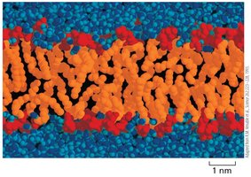
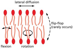
Fluid Nature of Lipid Bilayer
The lipid bilayer is a flexible, two-dimensional fluid, allowing rapid movement of lipids within each monolayer.
Lateral Diffusion: Lipids exchange places with neighbors, enabling rapid diffusion.
Rotation: Lipids rotate along their axis.
Flexion: Hydrocarbon tails flex and bend.
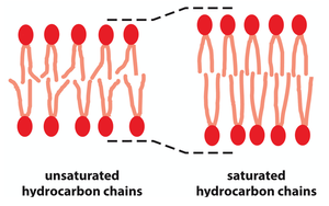
Factors Affecting Membrane Fluidity
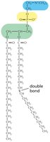
Membrane fluidity is influenced by phospholipid composition, hydrocarbon tail length, and degree of saturation.
Shorter Tails: Increase fluidity.
Unsaturated Tails: Contain double bonds, prevent tight packing, increase fluidity.
Saturated Tails: No double bonds, allow tight packing, decrease fluidity.
Role of Cholesterol in Membrane Fluidity
Animal cells modulate membrane fluidity by incorporating cholesterol, which has paradoxical effects depending on the surrounding lipids.
Cholesterol: Constrains motion of unsaturated fatty acid tails (decreases fluidity), but increases fluidity in saturated regions.
Membrane Assembly and Asymmetry
Phospholipid Synthesis and Distribution
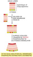
Membrane assembly begins in the endoplasmic reticulum (ER), where phospholipids are synthesized and inserted into the cytosolic half of the bilayer.
Scramblases: Enzymes that transfer phospholipids between bilayer halves, randomizing distribution.
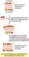
Asymmetric Distribution in Golgi and Plasma Membranes
The Golgi apparatus contains flippases and floppases, which selectively move specific phospholipids to maintain membrane asymmetry.
Flippases: Move phospholipids to cytosolic side (ATP-dependent).
Floppases: Move phospholipids to noncytosolic side (ATP-dependent).
Membrane Orientation and Protein Distribution
Membranes retain their orientation during vesicle transport, maintaining distinct inside and outside faces for both lipids and proteins.
Orientation: Essential for proper function of membrane proteins and signaling.
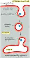
Specialized Functions: Different cells have distinct sets of membrane proteins, reflecting their specialized roles.
Membrane Proteins: Structure and Function
Role of Membrane Proteins
Membrane proteins constitute about 50% of the mass of animal cell plasma membranes and are responsible for most membrane functions.
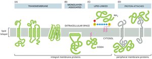
Types of Membrane Proteins
Membrane proteins associate with the lipid bilayer in several ways:
Transmembrane Proteins: Span the bilayer, amphipathic, can be single or multiple alpha helices, or beta barrels.
Monolayer-Associated Proteins: Anchored to one side by amphipathic alpha helix.
Lipid-Linked Proteins: Covalently attached to lipid molecules, entirely on one side.
Protein-Attached Proteins: Bound indirectly via noncovalent interactions with other membrane proteins.


Transmembrane Protein Structure
Polypeptide chains usually cross the lipid bilayer as an alpha helix, with hydrophobic side chains facing lipids and polar peptide bonds forming hydrogen bonds within the helix.
Alpha Helix: Maximizes hydrogen bonding, stabilizes protein within membrane.
Membrane Channels and Multipass Proteins
Multipass transmembrane proteins form aqueous pores, allowing small water-soluble molecules to cross the membrane.
Amphipathic Regions: Hydrophobic side chains face lipids, hydrophilic side chains face pore.
Solubilization and Study of Membrane Proteins
Membrane proteins can be solubilized using detergents, which disrupt hydrophobic interactions and bring proteins into solution.
Detergents: Hydrophobic ends interact with membrane proteins and lipids, hydrophilic ends solubilize proteins.
Example: Bacteriorhodopsin Structure and Function
Bacteriorhodopsin is a seven-transmembrane protein that acts as a proton pump, using a retinal chromophore to absorb light and transfer protons across the membrane.
Proton Pump: Transfers H+ from retinal to Asp 85, then across membrane, and takes up H+ from cytosol (Asp 96).
Membrane Reinforcement and Protein Mobility
Cell Cortex and Membrane Strengthening
The plasma membrane is reinforced by the cell cortex, a meshwork of fibrous proteins attached to the membrane by transmembrane proteins.
Spectrin: Main component in red blood cells, provides flexibility and strength.
Actin: Works with spectrin, attached via membrane proteins.
Mobility and Domains of Membrane Proteins
Many membrane proteins move freely within the bilayer, but some are confined to specific regions (membrane domains) by tethering or diffusion barriers.
Membrane Domains: Proteins can be tethered to cell cortex, extracellular matrix, or other cells.
Tight Junctions: Restrict protein diffusion, maintain domain integrity (e.g., in epithelial cells).
Cell Surface Carbohydrates
Glycoproteins and Glycocalyx
Many membrane proteins have covalently attached sugars, forming glycoproteins and proteoglycans. These carbohydrates face outward, creating a protective layer called the glycocalyx.
Glycoproteins: Short oligosaccharide chains.
Proteoglycans: Long polysaccharide chains.
Glycocalyx: Protects cell surface, aids in motility.
Cell-Cell Recognition and Adhesion
Cell-surface carbohydrates are crucial for cell-cell recognition and adhesion, especially in multicellular organisms.
Lectin Proteins: Bind to oligosaccharide side chains, mediating recognition.
Example: During bacterial infection, lectins on blood vessel walls recognize glycolipids and glycoproteins on neutrophils, enabling them to adhere and migrate out of the bloodstream.
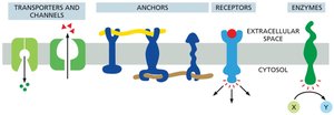
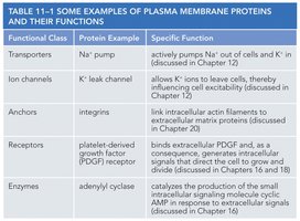
Table: Examples of Plasma Membrane Proteins and Their Functions
Functional Class | Protein Example | Specific Function |
|---|---|---|
Transporters | Na+ pump | Actively pumps Na+ out of cells and K+ in |
Ion channels | K+ leak channel | Allows K+ ions to leave cell, influencing cell excitability |
Anchors | Integrins | Link intracellular actin filaments to extracellular matrix proteins |
Receptors | Platelet-derived growth factor (PDGF) receptor | Binds extracellular PDGF and signals cell growth and division |
Enzymes | Adenylyl cyclase | Catalyzes production of cAMP, a small intracellular signaling molecule |
Summary
Cell membranes are complex, dynamic structures essential for cellular function, compartmentalization, communication, and recognition. Their composition, fluidity, and protein content are tightly regulated, enabling diverse biological processes central to genetics and cell biology. Additional info: The notes above expand on the original content with academic context, definitions, and examples to ensure completeness and clarity for genetics students.
