 Back
Study Guide: Mitosis and Meiosis (Concepts of Genetics, Chapter 2)
Study Guide - Smart Notes
 Tailored notes based on your materials, expanded with key definitions, examples, and context.
Cell Structure
Plasma Membrane, Cell Wall, and Glycocalyx
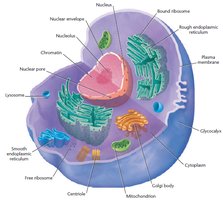
The plasma membrane forms the boundary of the cell and regulates the movement of substances in and out. In plants, the cell wall provides additional structural support, mainly composed of cellulose. Animal cells possess a glycocalyx, a covering of glycoproteins and polysaccharides, which contributes to the cell's biochemical identity and is under genetic control (e.g., blood group antigens, histocompatibility antigens).
Nucleus, Chromatin, Chromosomes, and Nucleolus
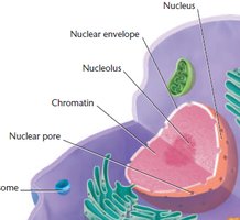
The nucleus is a membrane-bound organelle containing DNA complexed with proteins. Chromatin is the diffuse form of genetic material during interphase, while chromosomes are condensed structures visible during cell division. The nucleolus is the site of ribosomal RNA (rRNA) synthesis and ribosome assembly, with the nucleolus organizer region encoding rRNA.
Prokaryotic vs. Eukaryotic Cell Structure
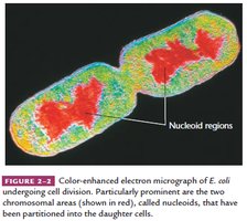
Prokaryotes lack a nuclear envelope and membranous organelles. Their DNA is circular and compacted in the nucleoid region, not associated with proteins as in eukaryotes. Prokaryotes do not have a nucleolus but contain genes for rRNA synthesis.
Cytoplasm and Organelles
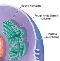
The cytoplasm contains organelles and cytosol, with microtubules and microfilaments forming the cytoskeleton for cell shape, motility, and organelle anchoring. The endoplasmic reticulum (ER) increases surface area for biochemical synthesis: smooth ER synthesizes fatty acids and phospholipids, while rough ER (with ribosomes) is the site of protein translation.
Mitochondria, Chloroplasts, and Centrioles
Mitochondria are responsible for ATP synthesis via oxidative respiration, while chloroplasts conduct photosynthesis in plants and algae. Both organelles contain their own DNA and replicate independently, supporting the endosymbiont hypothesis. Centrioles organize spindle fibers during mitosis and meiosis, facilitating chromosome movement.
Chromosomes Exist in Homologous Pairs in Diploid Organisms
Centromere and Chromosome Classification
The centromere is the constricted region of the chromosome, determining its appearance. Chromosomes are classified by centromere position:
Metacentric: Centromere in the middle
Submetacentric: Centromere between middle and end
Acrocentric: Centromere close to end
Telocentric: Centromere at the end
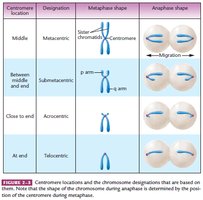
Homologous Chromosomes and Karyotype
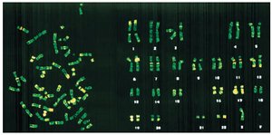
Somatic cells have a diploid number (2n) of chromosomes, with homologous pairs except for sex chromosomes. Karyotyping involves photographing and matching chromosomes. Sister chromatids are joined at a common centromere.
Genome, Locus, and Biparental Inheritance
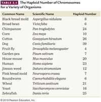
The haploid number is half the diploid number. The genome includes all genetic information in a haploid set. Homologous chromosomes have identical loci (gene sites), but alleles may differ. Biparental inheritance means one chromosome set from each parent.
Alleles and Sex Chromosomes
Alleles are alternative forms of the same gene. Sex chromosomes (X and Y) are not homologous in size or genetic content, but share homologous regions and behave as homologs during meiosis.
Mitosis Partitions Chromosomes into Dividing Cells
Purpose and Process of Mitosis
Mitosis is essential for asexual reproduction, growth, development, wound healing, and cell replacement. Genetic material is partitioned into two daughter cells during karyokinesis, followed by cytokinesis. Organelles replicate or are synthesized anew, resulting in two genetically identical cells.
Interphase and the Cell Cycle
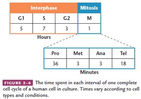
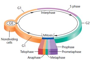
The cell cycle consists of division (mitosis) and nondivision (interphase) phases. Interphase includes G1 (cell growth), S (DNA synthesis), and G2 (preparation for mitosis). G1 is variable, S and G2 are consistent. Cells may enter G0 (quiescent stage) or continue cycling. Cancer cells bypass G0.
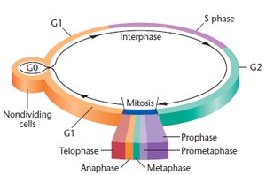
Cell Cycle Regulation and Checkpoints
Cell division cycle mutations (cdc) affect specific stages. Kinases and cyclins form cyclin-dependent kinases (CDKs), which phosphorylate target proteins to regulate the cycle. Major checkpoints:
G1/S checkpoint: Monitors cell size and DNA damage
G2/M checkpoint: Ensures DNA replication is complete and damage is repaired
M checkpoint: Monitors spindle fiber formation and attachment
Stages of Mitosis
Prophase
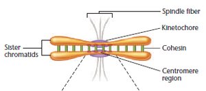
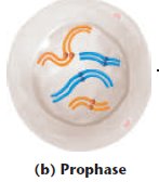
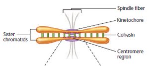
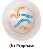
Centrioles migrate to opposite poles, organizing spindle fibers. Nuclear envelope and nucleolus disappear, chromatin condenses into chromosomes. Each chromosome forms two sister chromatids, held together by cohesin.
Prometaphase and Metaphase
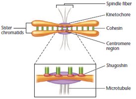
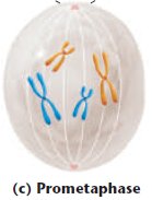
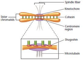
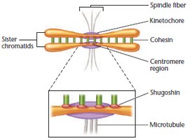
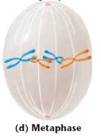
Chromosomes migrate to the metaphase plate. Spindle fibers attach to kinetochores. Cohesin is degraded by separase, except at the centromere, protected by shugoshin.
Anaphase
Sister chromatids separate (disjunction) and move to opposite poles, becoming daughter chromosomes. Migration is powered by motor proteins and ATP hydrolysis.
Telophase
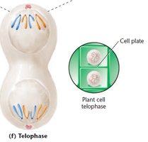
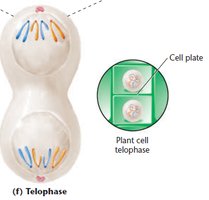
Chromosomes arrive at poles, cytokinesis completes. In plants, a cell plate forms; in animals, the cell membrane constricts. Chromosomes uncoil, nuclear envelope reforms, spindle fibers disappear.
Example Calculation
Diploid number = 16; chromatids at end of mitotic prophase = 32
Chromosomes moving to each pole during anaphase = 16
Meiosis Reduces Chromosome Number from Diploid to Haploid
Purpose and Genetic Variation
Meiosis reduces chromosome number by half, producing haploid gametes or spores. It ensures genetic continuity and increases genetic variation through crossing over and independent assortment.
Meiosis Process
Homologous chromosomes pair (synapsis), forming bivalents and then tetrads. Meiosis involves two divisions:
Meiosis I (Reductional): Homologs separate, reducing chromosome number
Meiosis II (Equational): Sister chromatids separate, maintaining chromosome number
Prophase I Stages
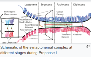
Leptonema: Chromatin condenses, chromosomes visible
Zygonema: Synaptonemal complex forms, homologs pair as bivalents
Pachynema: Synapsis completes, tetrads form
Diplonema: Chromatids separate except at chiasmata, crossing over occurs
Diakinesis: Chiasmata move toward ends, tetrads align at metaphase plate
Meiosis I: Metaphase, Anaphase, Telophase
Tetrads align randomly at metaphase plate. Cohesin is degraded except at centromere (protected by shugoshin). Homologs separate (disjunction); errors cause nondisjunction. Dyads (haploid number) are present at each pole.
Meiosis II: Prophase, Metaphase, Anaphase, Telophase
Dyads align at metaphase plate. Shugoshin is degraded, centromeres separate, sister chromatids move to opposite poles. Four haploid gametes result, each with unique genetic combinations.
Example Calculation
Diploid number = 16; tetrads in prophase I = 8
Dyads in prophase II = 8
Monads moving to each pole in anaphase II = 8
Development of Gametes: Spermatogenesis vs. Oogenesis
Spermatogenesis
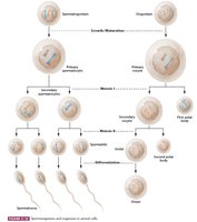
Occurs in testes. Diploid spermatogonium becomes primary spermatocyte, undergoes meiosis I to form secondary spermatocytes (haploid dyads), then meiosis II to form spermatids. Spermatids differentiate into motile sperm. Process can be continuous or periodic.
Oogenesis
Occurs in ovaries. Primary oocyte undergoes meiosis I, forming a secondary oocyte (most cytoplasm) and first polar body (little cytoplasm). Meiosis II produces an ootid and second polar body; ootid differentiates into mature ovum. In humans, meiosis I begins in embryonic ovary and arrests in prophase I, resuming years later. Meiosis II completes only after fertilization.
Summary Table: Key Differences in Gametogenesis
Process | Location | Outcome | Timing |
|---|---|---|---|
Spermatogenesis | Testes | Four sperm | Continuous/Periodic |
Oogenesis | Ovaries | One ovum, polar bodies | Arrested, resumes at ovulation/fertilization |
